You clone the full-length gene. Express it. Inclusion bodies. You try with tags. Still insoluble. You try insect cells. It expresses, but aggregates during purification. You truncate the C-terminus—guess where. The protein is now soluble but completely inactive.
Six months gone, and you still don't have a working construct.
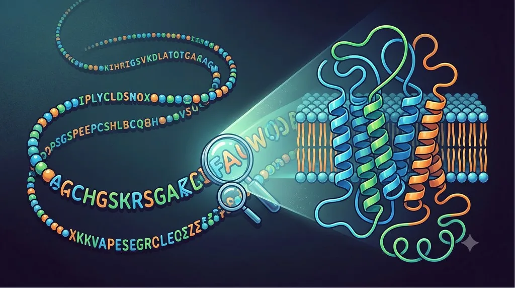
The problem isn't your expression system or purification protocol. It's your construct boundaries. Defining where a protein starts and ends—what to include, what to truncate—is the single most important decision in structural biology.
Key Takeaways
-
80-90% of construct failures are due to wrong boundaries
-
Main problems: Disordered termini (40%), flexible internal loops (20%), wrong domain boundaries (30%), bad tag placement (10%)
-
Average constructs tested: 5-15 per successful structure (traditional trial-and-error)
-
Cost of poor boundaries: $50-100K wasted per failed construct (6-12 months)
-
Modern solution: AI-driven boundary prediction reduces constructs tested from 10+ to 1-3
-
Success rate improvement: 60-80% first-construct success (vs 15-25% traditional)
What Are Construct Boundaries?
Construct boundaries define which residues you express and purify:
-
N-terminal boundary: Where does your protein start?
-
C-terminal boundary: Where does it end?
-
Internal boundaries: Which loops or domains do you include, truncate, or replace?
Example:
-
Full-length: Residues 1-450
-
Your construct: Residues 28-412 (N-terminal truncation of 27 residues, C-terminal truncation of 38 residues)
Why Boundaries Matter: The Goldilocks Principle
Include too much (disordered regions):
-
Flexible termini prevent crystallization
-
Disordered loops cause aggregation
-
PTMs create heterogeneity (blocks cryo-EM)
-
Result: Expresses but won't crystallize/freeze
Include too little (remove essential domains):
-
Protein loses stability (unfolds, aggregates)
-
Loses function (active site disrupted)
-
Result: Doesn't express, or is insoluble/inactive
Just right (structured core, minimal disorder):
-
Protein is stable, monodisperse
-
Crystallizes or forms good cryo-EM grids
-
Retains function
-
Result: Structure determination succeeds
Problem 1: Disordered N- and C-Terminal Tails (40% of Failures)
What it means: Your construct includes long (>15 residues) disordered regions at the termini.
Why It's a Problem
For crystallization:
-
Disordered residues adopt multiple conformations
-
Cannot form regular crystal lattice (requires identical packing)
-
Act as "entropy shields" (prevent crystal nucleation)
For cryo-EM:
-
Flexible termini create heterogeneity
-
2D class averages become fuzzy
-
Reduces resolution
For biochemistry:
-
Disordered termini contain protease cleavage sites
-
Protein degrades during purification
-
Stability decreases (lower Tm)
How to Diagnose
1. AlphaFold pLDDT (per-residue confidence):
-
Blue (pLDDT >90): Well-ordered
-
Green (pLDDT 70-90): Likely ordered
-
Orange (pLDDT 50-70): Questionable
-
Red (pLDDT <50): Disordered
Rule of thumb: If >15 consecutive residues have pLDDT <50 at N- or C-terminus → Truncate
2. Disorder prediction tools:
-
IUPred: Score >0.5 = disordered
-
PrDOS: Probability >0.5 = disordered
-
DISOPRED3: Binary prediction
3. Sequence composition:
-
Disordered regions enriched in: Gly, Ser, Pro, Glu, Lys, Gln
-
Structured regions enriched in: Trp, Phe, Tyr, Ile, Val, Leu, Cys
-
Calculate composition: >40% disorder-promoting → likely disordered
4. Homolog comparison:
-
Find crystal structures of homologs in PDB
-
Check what boundaries they used
-
Disordered termini often truncated in successful structures
Example: Bacterial Enzyme
Full-length (1-380):
-
AlphaFold pLDDT:
-
Residues 1-22: pLDDT 30-50 (disordered)
-
Residues 23-355: pLDDT 85-95 (structured)
-
Residues 356-380: pLDDT 40-60 (disordered)
-
Test constructs:
-
Construct 1 (1-380, full-length): Expresses, won't crystallize
-
Construct 2 (23-355, truncate both): Expresses, crystallizes, 2.3 Å
-
Construct 3 (30-350, aggressive): Expresses, lower yield/stability
Winner: Construct 2 (remove disorder, keep structured core)
Problem 2: Flexible Internal Loops (20% of Failures)
What it means: Long (>15 residues) flexible loops connecting structured domains.
Why It's a Problem
For crystallization:
-
Flexible loops adopt multiple conformations
-
Prevent ordered crystal packing
-
Often proteolytically cleaved during crystallization
For cryo-EM:
-
Loops create local disorder
-
Reduce resolution in those regions
-
May cause preferred orientation
Common Locations
GPCRs:
-
Intracellular loop 3 (ICL3): 20-80 residues, highly flexible
-
Extracellular loop 2 (ECL2): Can be long and flexible
Multi-domain proteins:
-
Linkers between domains (5-50 residues)
-
Hinge regions (allow domain movement)
Enzymes:
-
Active site loops (flexible when substrate-free)
-
Regulatory loops
How to Diagnose
1. AlphaFold pLDDT analysis:
-
Internal loops with pLDDT <70 are likely flexible
-
Long loops (>20 residues) with pLDDT 70-85 may still cause problems
2. B-factors in homolog structures:
-
High B-factors (>80 Ų) indicate flexibility
-
These regions will be problematic
3. Proteolysis sensitivity:
-
Limited proteolysis (trypsin 1:1000, 30 min at RT)
-
Flexible loops cleaved preferentially
-
Mass spec identifies cleavage sites
The GPCR ICL3 Problem
Famous example: β2-adrenergic receptor
-
ICL3 (residues ~230-270): 40 residues, highly flexible
-
Wild-type: Cannot crystallize
-
Solution: Replace ICL3 with T4 lysozyme
-
Construct: Residues 1-229 + T4L + 271-350
-
Result: First GPCR crystal structure (2007, Nobel Prize 2012)
Why it worked:
-
T4L is rigid, well-behaved
-
Provides crystal contacts
-
Acts as "fiducial marker" for alignment
Problem 3: Wrong Domain Boundaries (30% of Failures)
What it means: You've truncated too aggressively and removed essential structural elements.
Why It's a Problem
Protein destabilization:
-
Removing C-terminal helix exposes hydrophobic core
-
Protein unfolds, aggregates
-
Lower Tm, reduced stability
Loss of function:
-
Active site disrupted
-
Cofactor binding site incomplete
-
Regulatory domain missing
How to Diagnose
1. AlphaFold structure inspection:
-
Check if truncation cuts through secondary structure
-
Bad: Truncate in middle of helix or β-strand
-
Good: Truncate in loop between elements
2. Domain architecture prediction:
-
Pfam: Identifies conserved domains
-
InterPro: Comprehensive domain annotation
-
Rule: Don't truncate within a Pfam domain
3. Homolog comparison:
-
Align with PDB homologs
-
Check if successful structures include the region you're removing
4. Stability testing:
-
Express truncated construct, measure Tm
-
Compare to longer construct
-
If ΔTm < -5°C → removed something important
Case Study: Removing Essential Helix
Target: Novel bacterial enzyme
Attempt 1:
-
Construct: Residues 1-320 (removed C-terminal 30 residues)
-
Expression: Inclusion bodies (completely insoluble)
Analysis:
-
AlphaFold structure: Residues 310-330 form amphipathic helix
-
Helix packs against core (stabilizes hydrophobic pocket)
-
Removing it exposes core → aggregation
Attempt 2:
-
Construct: Residues 1-340 (keep helix, remove last 10 disordered)
-
Expression: Soluble, stable
-
Crystallization: Success, 2.1 Å structure
Lesson: Even one helix can be critical. Respect secondary structure boundaries.
Problem 4: Wrong Fusion/Tag Placement (10% of Failures)
What it means: Affinity tag (His-tag, GST, MBP) or fusion protein placed where it disrupts folding or function.
Why It's a Problem
Fusion blocks active site:
-
Tag near substrate binding site
-
Protein cannot bind substrate
-
Appears inactive
Fusion disrupts oligomerization:
-
Protein normally forms dimer
-
Tag at dimerization interface prevents assembly
-
Monomeric protein is unstable
Fusion prevents membrane insertion:
-
For membrane proteins, N-terminal signal peptide required
-
Tag at N-terminus blocks signal peptide
-
Protein not inserted, aggregates
How to Diagnose
1. Check AlphaFold structure:
-
Visualize where N- and C-termini are located
-
Are they buried or surface-exposed?
-
Are they near active site or oligomerization interface?
2. Homolog comparison:
-
Where are termini in crystal structures?
-
Are they accessible?
3. Test both termini:
-
Construct A: His-tag at N-terminus
-
Construct B: His-tag at C-terminus
-
Test expression, solubility, activity
Tag Placement Rules
Rule 1: Surface-exposed termini are best
-
If AlphaFold shows N-terminus on surface → N-terminal tag okay
-
If buried → try C-terminal tag
Rule 2: Avoid active sites
-
Check where substrate binds
-
Place tag on opposite side
Rule 3: Avoid oligomerization interfaces
-
If protein forms dimer, check interface
-
Place tag away from contact surface
Rule 4: Use cleavable tags
-
For crystallography/cryo-EM: Remove tag after purification
-
Use TEV protease site, PreScission site, or SUMO
Multi-Domain Proteins: Special Considerations
Example: Full-length (1-520)
-
Domain 1 (catalytic): Residues 45-280
-
Linker (flexible): Residues 281-310
-
Domain 2 (regulatory): Residues 311-490
-
C-terminal tail (disordered): Residues 491-520
Good construct options:
-
Construct A: 45-280 (catalytic domain only)
-
Construct B: 45-490 (both domains, no disordered tail)
-
Construct C: 311-490 (regulatory domain only)
Bad construct options:
-
Construct D: 45-250 (truncates within catalytic domain) → Unstable
-
Construct E: 100-490 (removes part of catalytic domain) → Inactive
Key principle: Keep entire domains, remove linkers between them.
Membrane Proteins: Transmembrane Boundaries
The Challenge
-
Need to include entire transmembrane (TM) helices
-
Truncating in middle of TM helix → unstable, aggregates
-
Including too much intracellular/extracellular domain → flexible, prevents crystallization
How to Define TM Boundaries
1. Predict TM helices:
-
TMHMM: Classic tool, reliable
-
DeepTMHMM: Deep learning-based, more accurate
-
Orbion: Integrates TM prediction with structure
2. AlphaFold structure:
-
TM helices have high pLDDT (usually >90)
-
TM regions form distinct bundle
3. Hydropathy plot:
- Kyte-Doolittle plot identifies hydrophobic regions (TM helices)
Boundary Rules for Membrane Proteins
Rule 1: Include complete TM helices
-
Don't truncate in middle of helix
-
Start/end in loop regions
Rule 2: Truncate flexible loops
-
Especially long loops (>20 residues)
-
GPCRs: ICL3 is classic target
Rule 3: Remove signal peptide
-
Signal peptide directs to ER
-
Not needed for recombinant expression
-
Predict with SignalP
Understanding Your Problem: Quick Diagnostic
| Symptom | Likely Problem | Quick Test |
|---|---|---|
| AlphaFold has long orange/red termini | Disordered termini | Check pLDDT <50 |
| Internal orange/red loops (>20 residues) | Flexible loops | Check pLDDT, compare homologs |
| Protein insoluble after truncation | Wrong domain boundary | Check if cut through helix/strand |
| Protein inactive after expression | Removed essential domain | Check Pfam, homolog structures |
| Low expression despite tag | Bad tag placement | Try opposite terminus |
| Membrane protein won't insert | Tag blocks signal peptide | Remove N-terminal tag |
Key Takeaway
Construct boundary design isn't guesswork. It's a systematic engineering problem with predictable failure modes:
-
Disordered termini (40%): Include flexible tails that prevent crystallization
-
Flexible loops (20%): Internal disorder blocks structure determination
-
Wrong domain boundaries (30%): Remove essential structural elements
-
Bad tag placement (10%): Tags disrupt function or folding
Understanding your boundary problem is the first step to fixing it.