In this article, we'll show you exactly how to design optimal boundaries before you clone.
The paradigm shift: Stop guessing. Start predicting.
Traditional approach: Clone full-length → Fail → Guess truncations → Fail → Test 10-15 constructs → Maybe succeed
Modern AI-driven approach: Analyze structure → Predict boundaries → Design 2-3 rational constructs → Test → Succeed
This guide provides the complete workflow, from sequence to optimized construct.
Key Takeaways
-
Design workflow: 6 systematic steps (sequence → boundaries → constructs → validation)
-
Success rate: 60-80% first-construct success with AI predictions (vs 15-25% traditional)
-
Time saved: 3-6 months per project (vs trial-and-error)
-
Tools: Free tools work (AlphaFold, IUPred), but Orbion reduces design time from 4-6 hours to 15 minutes
-
Fusion proteins: Use when native boundaries fail (GPCRs, unstable proteins)
-
Validation: Always predict Tm, check secondary structure integrity
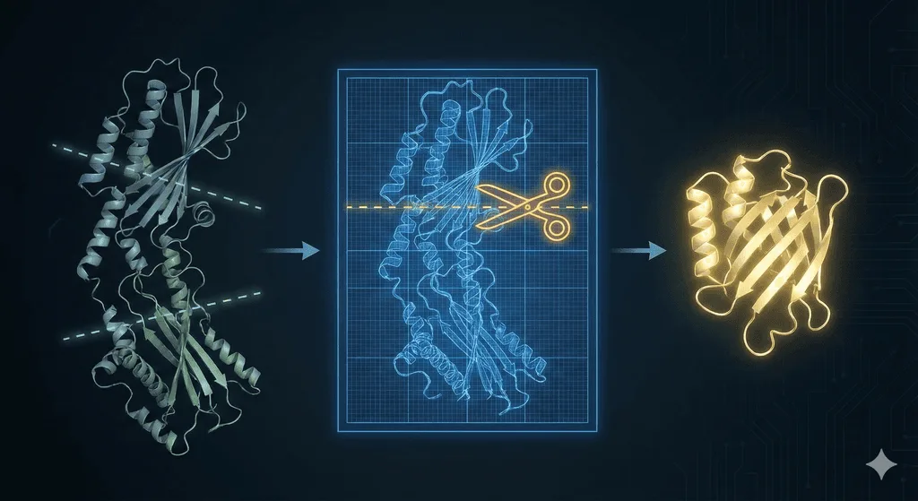
The 6-Step Design Workflow
Step 1: Get Your Structure Prediction (5 minutes)
Free approach:
-
Go to AlphaFold Database
-
Search by UniProt ID or sequence
-
Download structure (PDB file) and pLDDT JSON
Orbion approach:
-
Upload sequence to Orbion
-
Automatic structure prediction with integrated boundary analysis
-
Get annotated boundaries with confidence scores
What to look for:
-
pLDDT coloring: Blue/green = ordered, orange/red = disordered
-
Overall fold: Is protein single-domain or multi-domain?
-
Termini location: Buried or surface-exposed?
Red flags:
-
Long disordered termini (>20 residues, pLDDT <50)
-
Internal disordered loops (>15 residues, pLDDT <70)
-
Multi-domain with flexible linkers
Step 2: Predict Disorder and Flexibility (10 minutes)
Free tools:
IUPred3: https://iupred.elte.hu/
-
Paste sequence
-
Select "Long disorder" option
-
Score >0.5 = disordered
-
Identify continuous stretches >15 residues
DISOPRED3: http://bioinf.cs.ucl.ac.uk/psipred/
-
Paste sequence
-
Binary prediction (ordered/disordered)
-
Cross-check with IUPred
What to record:
-
N-terminal disorder: Residues X-Y (length, score)
-
C-terminal disorder: Residues X-Y (length, score)
-
Internal loops: Position, length, score
Example output:
Protein: 1-380
N-terminal disorder: 1-22 (22 residues, IUPred >0.6)
Structured core: 23-355 (pLDDT >85)
C-terminal disorder: 356-380 (25 residues, IUPred >0.5)
Step 3: Analyze Domain Architecture (10 minutes)
Free tools:
Pfam: https://pfam.xfam.org/
-
Paste sequence
-
Identifies conserved domains
-
Shows boundaries for each domain
InterPro: https://www.ebi.ac.uk/interpro/
-
Comprehensive domain annotation
-
Includes functional sites
Critical rules:
-
Don't truncate within a Pfam domain (unless you know what you're doing)
-
Keep complete secondary structure elements (don't cut helices/strands)
-
Truncate in loops between domains or elements
Example:
Full-length: 1-520
Pfam domains:
  - Kinase domain: 45-280
  - SH2 domain: 311-490
  - Disordered linker: 281-310
  - Disordered tail: 491-520

Valid constructs:
  ✓ 45-280 (kinase only)
  ✓ 311-490 (SH2 only)
  ✓ 45-490 (both domains, remove tail)
  ✗ 45-250 (truncates within kinase) → Unstable
  ✗ 100-490 (removes part of kinase) → Inactive
Step 4: Compare with Homolog Structures (15 minutes)
Why it matters: Successful structures in PDB show what boundaries actually work.
How to do it:
1. Find homologs:
-
Go to BLAST
-
Search against PDB database
-
Find homologs with >30% identity
2. Check boundaries used:
-
Open PDB entries
-
Note which residues were crystallized
-
Compare to full-length sequence
3. Identify consensus truncations:
-
If 3+ structures truncate N-terminus at residue ~25 → likely essential
-
If none include C-terminal 30 residues → likely disordered
Example: Bacterial enzyme homologs
| PDB ID | Identity | Construct Boundaries | Resolution |
|---|---|---|---|
| 1ABC | 45% | 28-350 (full-length 1-380) | 2.1 Å |
| 2DEF | 38% | 25-355 (full-length 1-385) | 1.8 Å |
| 3GHI | 52% | 30-348 (full-length 1-375) | 2.5 Å |
Consensus: All truncate N-terminus (~25-30), all truncate C-terminus (~350), none include 1-25 or 355+
Your construct: 25-355 (high confidence)
Step 5: Design Your Construct Set (30 minutes)
Strategy: Design 2-3 constructs with different risk levels
Construct A: Conservative (highest stability, lowest risk)
-
Remove only clearly disordered regions (pLDDT <50, IUPred >0.5)
-
Keep all secondary structure elements
-
Based on homolog consensus
Construct B: Optimal (balance stability and crystallizability)
-
Remove all disordered regions (pLDDT <70)
-
Truncate flexible loops if >20 residues
-
Most likely to succeed
Construct C: Aggressive (highest crystallizability, higher risk)
-
Minimal boundaries (only structured core)
-
May sacrifice some stability
-
Use if Construct B fails
Example design:
Target: Novel kinase (1-420)
-
AlphaFold pLDDT:
-
1-18: pLDDT 35-50 (disordered)
-
19-380: pLDDT 85-95 (structured)
-
381-420: pLDDT 40-60 (disordered)
-
-
Pfam: Kinase domain 25-375
-
Homologs: Truncate at ~20 and ~380
Construct set:
Construct A (Conservative): 19-385
  - Removes clear disorder (pLDDT <50)
  - Keeps some questionable C-terminus (pLDDT 50-60)
  - Risk: May not crystallize (some disorder remains)

Construct B (Optimal): 25-375
  - Aligns with Pfam domain
  - pLDDT >85 throughout
  - Matches homolog consensus
  - Risk: Lowest (recommended first)

Construct C (Aggressive): 30-370
  - Removes 5 extra residues each end
  - Maximum order (pLDDT >90)
  - Risk: May reduce stability
Which to test first: Construct B (optimal balance)
Step 6: Validate Your Design (10 minutes)
Before you order primers, check:
1. Secondary structure integrity:
-
Load AlphaFold structure in PyMOL or ChimeraX
-
Check N-terminal boundary: Does it cut through helix/strand?
-
Check C-terminal boundary: Same check
-
Rule: Truncate in loops, not secondary structure
2. Termini accessibility:
-
Are N- and C-termini surface-exposed?
-
If buried → fusion tag will cause problems
-
Plan tag placement accordingly
3. Active site check:
-
Visualize active site (if known)
-
Ensure your boundaries include all catalytic residues
-
Ensure fusion tag won't block substrate access
4. Predicted stability (Orbion only):
-
Orbion predicts Tm for each construct
-
Compare Construct A vs B vs C
-
If ΔTm >10°C → choose more stable construct
Free Tools vs Orbion: When to Use What
Free Tools Approach (Total: 4-6 hours)
Advantages:
-
Zero cost
-
Good for single constructs
-
Educational (you learn the details)
Workflow:
-
AlphaFold Database (5 min) → Structure
-
IUPred3 (10 min) → Disorder
-
Pfam (10 min) → Domains
-
BLAST + manual PDB inspection (30 min) → Homologs
-
Manual analysis in PyMOL (60 min) → Boundary design
-
Repeat for each construct variant (30 min each)
Total time: 4-6 hours for 3 constructs
Best for:
-
Students, academic labs
-
Single-protein projects
-
Learning structural biology principles
Orbion Approach (Total: 15 minutes)
Advantages:
-
Automated workflow
-
Integrated analysis (disorder + domains + homologs)
-
Predicted Tm for stability comparison
-
Batch processing (multiple constructs simultaneously)
-
Confidence scores for each boundary recommendation
Workflow:
-
Upload sequence (1 min)
-
Automatic boundary analysis (5 min)
-
Review 2-3 suggested constructs with Tm predictions (5 min)
-
Select optimal construct (2 min)
-
Export to ordering (2 min)
Total time: 15 minutes for full analysis
What Orbion adds:
-
AstraSTASIS: Predicts Tm for each construct (know stability before expressing)
-
Homolog mining: Automatic BLAST + PDB analysis
-
Boundary confidence: Statistical scoring based on multiple predictors
-
Tag optimization: Suggests N- vs C-terminal placement
-
Batch mode: Analyze 10+ constructs in parallel
Best for:
-
Biotech, pharma (time = money)
-
Multi-construct screening
-
High-throughput projects (>5 proteins/month)
-
When first-construct success is critical
Cost-benefit:
-
Saves 4-5 hours per protein × $100/hour scientist = $400-500 saved
-
Orbion cost: Starts at $99/month (unlimited analyses)
-
Break-even: 1 protein per month
When to Use Fusion Proteins
Sometimes native boundaries aren't enough. Fusion proteins can rescue difficult targets.
Fusion Type 1: Solubility Tags (MBP, GST, SUMO)
When to use:
-
Protein expresses but insoluble
-
Even with optimized boundaries
Common tags:
MBP (Maltose Binding Protein, 42 kDa):
-
Best for: Solubilizing aggregation-prone proteins
-
Placement: N-terminal
-
Cleavage: TEV protease site
-
Success rate: 70-80% of insoluble proteins become soluble
GST (Glutathione S-Transferase, 26 kDa):
-
Best for: Small proteins (<20 kDa)
-
Placement: N-terminal
-
Cleavage: PreScission protease
-
Bonus: Forms dimer (stabilizes small proteins)
SUMO (Small Ubiquitin-like Modifier, 12 kDa):
-
Best for: Eukaryotic proteins
-
Placement: N-terminal
-
Cleavage: SUMO protease (leaves no extra residues)
-
Bonus: Enhances expression in E. coli
Design rule:
-
Always include protease cleavage site
-
Remove tag after purification for crystallography/cryo-EM
-
Test activity with and without tag
Fusion Type 2: Crystallization Helpers (T4L, BRIL)
When to use:
-
Protein soluble but won't crystallize
-
Especially for membrane proteins (GPCRs)
T4 Lysozyme (T4L, 18 kDa):
-
Famous use: β2-adrenergic receptor (2007 Nobel Prize)
-
Strategy: Replace flexible ICL3 with T4L
-
Why it works:
-
Rigid, well-behaved protein
-
Provides crystal contacts
-
Acts as fiducial marker for cryo-EM alignment
-
BRIL (Thermostable apocytochrome b562, 11 kDa):
-
Use: GPCR crystallization
-
Advantage: Smaller than T4L (less disruption)
-
Placement: Replace ICL3 or insert at N-terminus
Design strategy:
GPCR (1-350):
  - TM1-TM7: Residues 25-320
  - ICL3 (flexible): Residues 230-270 (40 residues)

Fusion construct:
  - Residues 25-229 (TM1-TM5)
  - T4L insertion (replaces ICL3)
  - Residues 271-320 (TM6-TM7)
  - Remove C-terminal tail (321-350)

Result: Rigid, crystallizable GPCR
Fusion Type 3: Nanobodies/DARPins (Conformational Stabilization)
When to use:
-
Protein is dynamic (multiple conformations)
-
Need to lock specific conformation for structure
How it works:
-
Co-express protein + nanobody
-
Nanobody binds, stabilizes one conformation
-
Reduces conformational heterogeneity
-
Easier to crystallize or freeze for cryo-EM
Example: G protein-coupled receptor
-
GPCR has active and inactive states
-
Nanobody binds active state
-
Locks conformation
-
Enables structure determination of active state
Orbion advantage:
-
AstraBIND: Predicts nanobody epitopes
-
Design nanobodies targeting specific conformations
Case Study: Multi-Domain Protein Optimization
Target: Novel bacterial enzyme with regulatory domain
Full-length: 1-520
-
N-terminal tail: 1-44 (disordered)
-
Catalytic domain: 45-280 (structured)
-
Linker: 281-310 (flexible)
-
Regulatory domain: 311-490 (structured)
-
C-terminal tail: 491-520 (disordered)
Traditional Approach (Failed)
Attempt 1: Full-length (1-520)
-
Expression: Low yield
-
Purification: Multiple degradation bands
-
Crystallization: No crystals after 300 conditions
-
Result: Failure (disordered tails cause problems)
Attempt 2: Remove tails (45-490)
-
Expression: Good
-
Purification: Clean, but some degradation
-
Crystallization: No crystals
-
Analysis: Limited proteolysis shows cleavage in linker (281-310)
-
Result: Partial success (linker still flexible)
Total time: 8 months, 2 failed attempts
AI-Driven Approach (Success)
Step 1: AlphaFold analysis
-
N-terminal 1-44: pLDDT 30-50 (remove)
-
Catalytic domain: pLDDT 90-95 (keep)
-
Linker: pLDDT 50-70 (problem area)
-
Regulatory domain: pLDDT 85-92 (keep)
-
C-terminal 491-520: pLDDT 35-50 (remove)
Step 2: Homolog check
-
Found 5 homologs in PDB
-
3 structures: Catalytic domain only (45-280)
-
2 structures: Regulatory domain only (311-490)
-
None: Full two-domain construct
-
Conclusion: Domains likely independent, linker prevents co-crystallization
Step 3: Construct design
Construct A: 45-280 (catalytic domain only)
  - Remove N-terminal tail, linker, regulatory domain, C-terminal tail
  - Pfam: Complete catalytic domain
  - Risk: May lose regulatory function

Construct B: 311-490 (regulatory domain only)
  - For understanding regulation mechanism separately

Construct C: 45-490 + linker replacement
  - Replace flexible linker (281-310) with rigid GGGGS linker
  - Risk: May disrupt domain-domain communication
Step 4: Testing
Construct A (45-280):
-
Expression: Excellent (50 mg/L)
-
Purification: Clean, monodisperse
-
Crystallization: Success (2.1 Å structure in 2 weeks)
-
Activity: 60% of full-length (good enough for structural studies)
-
Result: Success
Construct B (311-490):
-
Expression: Good
-
Crystallization: Success (2.5 Å)
-
Result: Second structure (regulatory mechanism revealed)
Total time: 6 weeks for 2 structures (vs 8 months with traditional)
Key lesson: Sometimes splitting multi-domain proteins is better than trying to crystallize them together.
Membrane Proteins: Special Workflow
Membrane proteins require additional considerations.
Extra Steps for Membrane Proteins
1. Predict transmembrane helices:
DeepTMHMM: https://dtu.biolib.com/DeepTMHMM
-
Predicts TM helix positions
-
Identifies inside/outside orientation
Output:
Protein: 1-450
Signal peptide: 1-25 (remove)
TM1: 35-55
TM2: 70-90
TM3: 110-130
...
TM7: 310-330
C-terminal tail: 331-450 (cytoplasmic, disordered)
2. Define TM boundaries:
Rules:
-
Include complete TM helices (don't truncate in middle)
-
Start boundary 5-10 residues before first TM helix
-
End boundary 5-10 residues after last TM helix
-
Remove signal peptide (not needed for recombinant expression)
-
Truncate long intracellular/extracellular loops
3. Handle flexible loops:
GPCRs: Intracellular loop 3 (ICL3) is usually 20-80 residues, highly flexible
Options:
-
Option A: Remove ICL3 entirely (if not functionally critical)
-
Option B: Replace with short linker (GGGGS)
-
Option C: Replace with T4 lysozyme (gold standard for crystallization)
Example construct:
GPCR (1-350):
Native: TM1-ICL1-TM2-ECL1-TM3-ICL2-TM4-ECL2-TM5-ICL3-TM6-ECL3-TM7

Optimized construct:
  - Remove signal peptide (1-25)
  - TM1-TM5: 26-229
  - Replace ICL3 with T4L
  - TM6-TM7: 271-320
  - Remove C-terminal tail (321-350)

Final: 26-229-T4L-271-320
Pre-Cloning Checklist
Before you order primers, verify:
Boundary Checklist
-
[ ] Disordered termini removed: pLDDT <50 regions truncated
-
[ ] Secondary structure intact: No cuts through helices/strands
-
[ ] Domain boundaries respected: Pfam domains complete
-
[ ] Homolog consensus: Boundaries match successful structures
-
[ ] Flexible loops assessed: Internal disorder identified
-
[ ] Active site included: All catalytic residues present
-
[ ] Termini accessible: N/C-termini surface-exposed (for tags)
Tag Design Checklist
-
[ ] Tag placement chosen: N- or C-terminal (based on structure)
-
[ ] Cleavage site included: TEV, PreScission, or SUMO protease
-
[ ] Tag won't block active site: Checked in structure viewer
-
[ ] Tag won't block oligomerization: Checked dimer interface
Expression Design Checklist
-
[ ] Codon optimized: For expression host (E. coli, insect, mammalian)
-
[ ] Rare codons avoided: Check codon usage
-
[ ] Signal peptide handled: Removed (if not needed) or kept (if required)
-
[ ] PTMs considered: Express in appropriate system (mammalian for glycosylation)
Construct Set Checklist
-
[ ] 2-3 variants designed: Conservative, optimal, aggressive
-
[ ] Boundaries differ by 5-10 residues: Small variations to test
-
[ ] Stability estimated: Predicted Tm for each (if using Orbion)
-
[ ] Testing order decided: Start with optimal construct
Common Mistakes to Avoid
Mistake 1: Not Checking Homologs
Problem: You design boundaries based only on disorder prediction, ignore PDB
Result: Your boundaries don't match proven successful constructs
Fix: Always BLAST against PDB, check what worked for homologs
Mistake 2: Cutting Through Secondary Structure
Problem: You truncate at residue 250 because it's "round number"
Result: You cut through C-terminal helix, protein unstable
Fix: Always visualize structure, truncate in loops only
Mistake 3: Removing Too Much
Problem: AlphaFold shows pLDDT 60-70 at termini, you remove it
Result: Protein less stable (removed stabilizing element)
Fix: Be conservative with "gray zone" (pLDDT 60-80), test longer construct first
Mistake 4: Ignoring Tag Placement
Problem: You always put His-tag at N-terminus (habit)
Result: Tag blocks active site, protein appears inactive
Fix: Check structure, place tag away from functional sites
Mistake 5: Not Testing Multiple Constructs
Problem: You design one "perfect" construct
Result: It fails, you don't have backup
Fix: Always design 2-3 variants, test in parallel
Advanced: When Standard Boundaries Fail
Problem: All Constructs Aggregate
You've tried:
-
Optimal boundaries (removed disorder)
-
Conservative boundaries (kept more)
-
Aggressive boundaries (minimal)
-
All aggregate during expression or purification
Solutions:
1. Try fusion tags (MBP, GST, SUMO):
-
MBP most effective for aggregation
-
Express as MBP-fusion, cleave after purification
2. Change expression system:
-
If E. coli fails → Try insect cells (Sf9/High Five)
-
If insect fails → Try mammalian (HEK293, CHO)
3. Lower expression temperature:
-
Express at 18°C instead of 37°C
-
Slower folding, less aggregation
4. Add chaperone co-expression:
-
GroEL/GroES for E. coli
-
Helps proper folding
5. Screen for stabilizing mutations (Orbion):
-
AstraSTASIS predicts stabilizing point mutations
-
Increase Tm by 5-15°C
-
More stable = less aggregation
Problem: Protein Soluble But Inactive
You've truncated boundaries, protein expresses and purifies, but has no activity
Diagnosis:
1. Check what you removed:
-
Did you remove part of active site?
-
Did you remove regulatory domain needed for activity?
-
Did you remove cofactor binding site?
2. Compare with full-length:
-
Express longer construct (more conservative boundaries)
-
Test activity
-
If active → you removed something essential
Solutions:
1. Extend boundaries:
-
Add back 10-20 residues at each terminus
-
Test which end matters
2. Include regulatory domain:
-
If multi-domain, include both catalytic + regulatory
-
Accept that you may need fusion approach for crystallization
3. Add back cofactor:
-
Some proteins need metal ions (Zn²⁺, Mg²⁺) or organic cofactors
-
Supplement during expression/purification
Key Takeaway
Construct boundary design is systematic engineering, not guesswork:
The 6-step workflow:
-
Get structure prediction (AlphaFold)
-
Predict disorder (IUPred, DISOPRED)
-
Analyze domains (Pfam, InterPro)
-
Compare homologs (BLAST + PDB)
-
Design 2-3 constructs (conservative/optimal/aggressive)
-
Validate before cloning (check secondary structure, termini)
Tools:
-
Free: 4-6 hours, good for learning
-
Orbion: 15 minutes, integrated workflow, Tm prediction
When to use fusions:
-
Solubility issues → MBP, GST, SUMO
-
Crystallization issues → T4L, BRIL
-
Conformational heterogeneity → Nanobodies
Success rate:
-
Traditional (trial-and-error): 15-25% first-construct success
-
AI-driven (predicted boundaries): 60-80% first-construct success
The paradigm shift is here. Stop guessing. Start predicting.