You cloned your antibody fragment into a vector you already had on the bench, kept the signal peptide that came with it, and ran the expression. The protein stayed inside the cell. Or it reached the periplasm with five extra residues hanging off the N-terminus. Or the supernatant titer was a tenth of what your colleague gets with the "same" construct in a different host.
Signal peptide selection is one of the highest-leverage decisions in a secretory expression run, and it's also one of the most frequently made on autopilot. The wrong leader can cost you an order of magnitude in yield — and signal peptide swaps that don't touch the mature protein at all have turned undetectable secretion into the dominant product in mammalian head-to-head screens (Güler-Gane et al., 2016).
Key Takeaways
- Signal peptides are not portable across hosts: a leader optimized for E. coli periplasmic export will not work in CHO, and vice versa — the translocation machinery, codon context, and cleavage enzymes differ.
- Two bacterial export routes exist: the Sec pathway moves unfolded chains and handles the great majority of secreted proteins; the Tat pathway (signal: TorA) moves already-folded proteins and is the right choice when your cargo folds in the cytoplasm or needs a cofactor.
- The signal-peptide/cargo pairing matters more than the "best" signal peptide: secretion efficiency is a property of the leader and the mature protein together, which is why empirical screening of several leaders beats picking one from a paper.
- Cleavage follows the (-3,-1) rule: signal peptidase needs small, uncharged residues at positions -1 and -3 relative to the cut site; violate it and you get uncleaved or mis-cleaved product even when the protein reaches the right compartment.
- Most poor-secretion phenotypes are diagnosable: cytoplasmic retention, incomplete cleavage, and post-translocation misfolding each leave a distinct signature, and each has a different fix.
Why the Signal Peptide Is the Gatekeeper
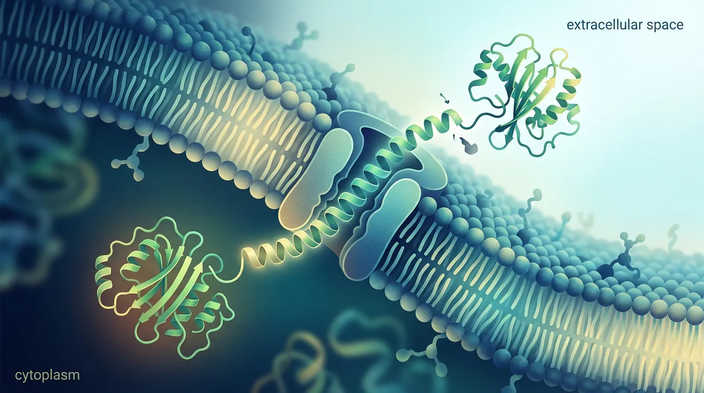
A signal peptide is a short N-terminal sequence — typically 15–30 residues — that routes a nascent protein into a secretory pathway and is cleaved off during or after translocation. Despite low sequence conservation, almost all signal peptides share the same three-part architecture: a positively charged n-region, a hydrophobic h-region that inserts into the membrane, and a polar c-region that presents the cleavage site to signal peptidase (Owji et al., 2018).
That architecture is universal. The specific sequences that work are not. A leader is read by host-specific machinery — the SRP and SecA/SecYEG translocon in bacteria, the SRP/Sec61 system in eukaryotes — and cleaved by host-specific peptidases. Swap the host and you change every one of those readers at once. This is why the single most common signal-peptide mistake is treating the leader as a portable part you carry over from whatever vector you happened to start with.
The core tension: the signal peptide that maximizes secretion is not a fixed property of the leader. It's a property of the leader paired with your specific cargo in your specific host. There is no universal best signal peptide, which is exactly why the field has moved toward screening small panels rather than betting on one sequence (Freudl, 2018).
The Two Bacterial Pathways: Sec vs Tat
In E. coli, two routes cross the inner membrane, and your signal peptide chooses which one.
The Sec Pathway (Default)
The Sec pathway transports proteins in an unfolded state and handles over 90% of secreted proteins in bacteria (Freudl, 2018). The unfolded chain is pushed through the SecYEG channel either post-translationally (the SecB route, used by most classical leaders like pelB and OmpA) or co-translationally (the SRP route, used by the DsbA leader). Once in the periplasm, the protein folds — and that's where the oxidizing environment and Dsb chaperones let disulfide-bonded proteins like antibody fragments mature correctly.
Use Sec when: your protein can fold after translocation, needs the periplasmic oxidizing environment for disulfides, or is a standard secretory target. This covers most scFv, Fab, and small enzyme work.
The Tat Pathway (Folded Cargo)
The twin-arginine translocation (Tat) pathway moves proteins that are already folded in the cytoplasm (Freudl, 2018). Its signal peptides carry a conserved twin-arginine (S/T-R-R-x-F-L-K) motif in the n-region — the TorA leader is the standard choice. Tat has a built-in quality-control feature: it generally rejects misfolded chains, so what reaches the periplasm tends to be correctly folded.
Use Tat when: your protein folds rapidly in the cytoplasm and resists Sec export, requires a cytoplasmically loaded cofactor (metal centers, FeS clusters), or assembles before export. The tradeoff is lower throughput — Tat is slower and lower-capacity than Sec.
Diagnostic question: Does your protein fold or pick up a cofactor before it can be exported? If yes, force it down Tat with TorA. If it can travel unfolded, stay on Sec.
Host-Specific Signal Peptides (and Why They Don't Travel)
Here's the rule that saves the most grief: a signal peptide is qualified for one host, not for a protein. The leaders below are grouped by the only context in which they work.
E. coli (periplasmic secretion)
- pelB — the Erwinia pectate lyase B leader; the default workhorse for antibody fragments and small proteins, post-translational Sec via SecB.
- OmpA — outer-membrane protein A leader; similar profile to pelB, often interchangeable, sometimes higher-yielding depending on cargo.
- PhoA — alkaline phosphatase leader; classic and well characterized, but rapidly folding passengers can fold before export and get stuck (Schierle et al., 2003).
- DsbA — directs co-translational export via the SRP pathway, exporting the chain before it has a chance to fold in the cytoplasm. This is the leader to reach for when a fast-folding protein resists the post-translational leaders (Schierle et al., 2003).
- TorA — the Tat leader, for folded cargo (see above).
Mammalian (CHO, HEK293 — secreted into medium)
- IgK (immunoglobulin kappa light-chain leader) — the de facto standard for secreted antibodies and Fc fusions; the benchmark other leaders are measured against.
- tPA (tissue plasminogen activator leader) — strong for many therapeutic proteins, but cargo-dependent: in at least one comparison it failed to secrete SEAP even with an inserted spacer, while CD33 and IgK restored secretion (Güler-Gane et al., 2016).
- CD33 — a heterologous leader that restored SEAP secretion (with an added downstream spacer) where the native tPA and a consensus leader failed in CHO (Güler-Gane et al., 2016).
- IL-2 — interleukin-2 leader, another well-validated mammalian secretory option.
Insect (Sf9, Hi5 — baculovirus)
- gp64 — baculovirus envelope glycoprotein leader; native to the system and a reliable default.
- melittin — the honeybee melittin leader; fusing it to a passenger drove over 5-fold more secreted product than a heterologous plant leader in insect cells (Tessier et al., 1991).
Yeast (Pichia pastoris, S. cerevisiae)
- alpha-MF — the S. cerevisiae α-mating-factor prepro leader; the most common and successful secretion signal for yeast. It's processed in stages: signal peptidase removes the pre-region in the ER, then the Kex2 endopeptidase cleaves the pro-region in the Golgi, and Ste13 trims the Glu-Ala spacer. The Kex2 cleavage context is itself tunable — modifying the P1′ residue has raised secreted yields of G-CSF in P. pastoris (Aggarwal & Mishra, 2021).
Why none of these travel: put IgK in front of a protein in E. coli and the bacterial SRP/Sec machinery doesn't recognize it the way mammalian SRP/Sec61 does, the codon context is wrong, and the bacterial signal peptidase cleavage preference differs. Put pelB into CHO and you get the mirror-image failure. The architecture is conserved; the recognition is not.
Getting the Cleavage Site Right: The (-3,-1) Rule
Reaching the right compartment is only half the job. If signal peptidase can't cut cleanly, you ship a product with a ragged or extended N-terminus — a real problem for therapeutics, where N-terminal heterogeneity is a release-spec headache.
Signal peptidase I recognizes a pattern, not a fixed sequence. The governing constraint is the (-3,-1) rule: positions -1 and -3 relative to the cleavage site must be small, uncharged residues (Ala is the canonical -1; the -3 position tolerates more variety but excludes large and charged side chains) (von Heijne, 1986). The c-region presents this motif to the peptidase. Break the rule — for instance by an awkward junction where your mature protein's first residues clash with the leader's c-region — and you get incomplete or shifted cleavage even when translocation itself succeeds.
Two practical consequences:
- The junction is part of the design. The first few residues of your mature protein influence cleavage. A leader that cleaves perfectly in front of one cargo can mis-cleave in front of another. This is another face of signal-peptide/cargo matching.
- Predict before you clone. Modern predictors model all five signal-peptide classes and flag the likely cleavage site directly from sequence (Teufel et al., 2022). Running your assembled construct through cleavage-site prediction takes minutes and catches junctions that would otherwise cost you a round of expression.
Troubleshooting Poor Secretion
When secretion underperforms, the failure almost always falls into one of three buckets. Each has a distinct signature.
1. Cytoplasmic Retention (the protein never left)
Signature: Protein is abundant in the cell lysate / cytoplasmic fraction but nearly absent from the periplasm or medium. Often appears in inclusion bodies.
Causes and fixes:
- Wrong pathway. A fast-folding cargo on a post-translational Sec leader folds before export. Switch to the co-translational DsbA leader, or route folded cargo through Tat/TorA (Schierle et al., 2003).
- Translocon saturation. Over-strong induction floods the Sec machinery. Lower the induction temperature, reduce inducer, or use a weaker promoter (Low et al., 2013).
- Poor leader/cargo match. Screen 3–4 alternative leaders for the host; secretion is an emergent property of the pair, not the leader alone.
2. Incomplete or Incorrect Cleavage (ragged N-terminus)
Signature: Protein is in the right compartment, but mass spec or Edman shows extra N-terminal residues, a mix of cleaved and uncleaved forms, or a shifted cut.
Causes and fixes:
- (-3,-1) violation at the junction. Re-examine the c-region/mature-protein boundary and adjust the spacer so positions -1 and -3 satisfy the rule (von Heijne, 1986).
- Yeast-specific Kex2 issues. With alpha-MF, ragged ends and Glu-Ala carryover trace to Kex2/Ste13 processing; tuning the Kex2 P1′ context resolves many cases (Aggarwal & Mishra, 2021).
- Verify in silico first. Predict the cleavage site for the exact assembled sequence before re-cloning (Teufel et al., 2022).
3. Post-Translocation Misfolding (secreted but dead/aggregated)
Signature: Protein reaches the medium or periplasm but is inactive, aggregated, or low-yield because it never folded correctly after export.
Causes and fixes:
- Disulfide-dependent protein in a non-oxidizing route. Antibody fragments need the periplasmic oxidizing environment; make sure you're on a Sec leader that delivers there, and co-express Dsb chaperones if needed.
- Cargo intrinsically aggregation-prone. No leader fixes a sticky mature sequence. Address aggregation hotspots and disorder in the protein itself before blaming the leader.
Diagnostic question: Is the protein somewhere it shouldn't be (retention), the right size plus a bit (cleavage), or the right place but non-functional (folding)? That single distinction points you straight at the right bucket.
Decision Tree: Picking a Signal Peptide
START: Which host?
│
├─ E. coli (periplasm) ─────────────────────────────────────────────
│   │
│   ├─ Does the protein fold / load a cofactor BEFORE export?
│   │     └─ Yes → TorA (Tat pathway)
│   │
│   ├─ Is it a fast-folding cargo that resists export?
│   │     └─ Yes → DsbA (co-translational SRP/Sec)
│   │
│   └─ Standard secretory target (scFv/Fab/small enzyme)?
│         └─ Screen pelB AND OmpA (and PhoA as a third); pick the winner
│
├─ Mammalian (CHO / HEK293) ─────────────────────────────────────────
│   └─ Antibody / Fc fusion / secreted therapeutic?
│         └─ Start with IgK; screen tPA, CD33, IL-2 in parallel
│            (tPA is cargo-dependent — don't assume it works)
│
├─ Insect (Sf9 / Hi5, baculovirus) ──────────────────────────────────
│   └─ gp64 (native default) OR melittin (often higher-yielding)
│
└─ Yeast (Pichia / S. cerevisiae) ───────────────────────────────────
    └─ alpha-MF prepro leader
       └─ Ragged N-terminus? Tune the Kex2 P1′ cleavage context

ALWAYS: predict the cleavage site for the FINAL assembled sequence
        before cloning, and verify the (-3,-1) rule at the junction.
Host-by-Host Selection Table
| Host | First-choice leader | Alternatives to screen | Pathway / mechanism | Watch for |
|---|---|---|---|---|
| E. coli (periplasm) | pelB | OmpA, PhoA | Sec, post-translational | Cytoplasmic retention of fast folders |
| E. coli (fast-folding cargo) | DsbA | — | Sec, co-translational (SRP) | Translocon saturation at high induction |
| E. coli (folded/cofactor cargo) | TorA | — | Tat (folded transport) | Lower throughput than Sec |
| CHO / HEK293 | IgK | tPA, CD33, IL-2 | SRP / Sec61, secreted to medium | tPA secretion is cargo-dependent |
| Sf9 / Hi5 (baculovirus) | gp64 | melittin | Insect secretory pathway | melittin often beats native leaders |
| Pichia / S. cerevisiae | alpha-MF (prepro) | — | Sec + Kex2/Ste13 processing | Glu-Ala carryover, Kex2 mis-cleavage |
Case Study: An scFv That Wouldn't Leave the Cytoplasm
Problem: A team expressing a single-chain antibody fragment in E. coli with the pelB leader saw strong total expression on a whole-cell gel but almost nothing in the periplasmic fraction. The protein was partitioning into inclusion bodies inside the cell.
Analysis: Total protein was high, so transcription and translation were fine — the failure was translocation. The scFv folded quickly in the cytoplasm, and pelB's post-translational Sec route can't export a chain that has already folded (Schierle et al., 2003). This is the classic cytoplasmic-retention signature: lots of protein, wrong compartment.
Solution: Two changes. First, swap pelB for the DsbA leader to force co-translational export via SRP, moving the chain across the membrane before it could fold. Second, drop the induction temperature and reduce inducer to avoid saturating the Sec translocon at the new higher export flux (Low et al., 2013). The assembled construct was run through cleavage-site prediction to confirm a clean (-3,-1) junction before re-cloning.
Outcome: Periplasmic, correctly disulfide-bonded scFv went from undetectable to the dominant product, with cleavage confirmed homogeneous by intact mass. No change was made to the mature antibody sequence — the entire fix was in the leader choice and the export conditions around it.
Pre-Cloning Checklist
Before you order DNA for a secretory construct, verify:
- Host matched. The signal peptide is qualified for your expression host, not carried over from a previous vector.
- Pathway matched. Sec for unfolded/disulfide cargo; Tat (TorA) for folded or cofactor-loaded cargo in E. coli.
- Panel chosen. You have at least 2–3 host-appropriate leaders queued to screen, not a single bet.
- Junction checked. Positions -1 and -3 relative to the predicted cut site are small and uncharged.
- Cleavage predicted. You ran the final assembled sequence (leader + mature protein) through cleavage-site prediction.
- Codon context handled. The leader's DNA is codon-optimized for the host, not just copied from the source organism.
- Assembly order correct. Signal peptide is at the extreme N-terminus, ahead of any N-terminal tag or fusion.
The Economics of Screening vs Guessing
| Approach | Up-front effort | Typical yield outcome | Real cost |
|---|---|---|---|
| Reuse whatever leader is in the vector | None | Hit-or-miss; often 5–10× below optimum | Weeks lost per failed round, repeated re-cloning |
| Pick one "best" leader from a paper | Low | Better, but cargo-dependent — the paper's cargo isn't yours | One re-clone when the pair underperforms |
| Screen a small host-matched panel (3–4 leaders) | Moderate (parallel constructs) | Consistently finds a near-optimal pair; large secretion gains are common | One expression round; pays back immediately |
ROI consideration: A signal-peptide swap changes nothing about your mature protein, your assay, or your purification — yet it routinely moves productivity substantially in mammalian antibody production (Kober et al., 2013) and over 5-fold in insect-cell secretion (Tessier et al., 1991). Parallelizing three or four leaders in one cloning round is the cheapest order-of-magnitude you'll find in a secretory campaign.
Bottom Line
Signal peptides are host-specific, cargo-dependent gatekeepers — not portable parts. Match the leader to your host, match the pathway (Sec vs Tat) to whether your protein folds before export, screen a small panel rather than betting on one sequence, and verify the (-3,-1) cleavage junction in silico before you clone.
How Orbion Helps
Most signal-peptide failures happen at the bench, but they're decided at the design stage — the moment you assemble leader, tags, linkers, and target into a construct. Orbion's Design tab (Beta) puts that decision on solid ground before any DNA is ordered.
The Design tab's component library includes a curated set of 14 signal peptides with verified sequences — every leader discussed above (pelB, OmpA, PhoA, DsbA, IgK, tPA, CD33, IL-2, gp64, melittin, alpha-MF, TorA) plus additional options like the Gaussia luciferase and Ost1 leaders — so you're choosing from validated sequences rather than copy-pasting from a vector map. When you build a construct manually or via AI generation, Orbion enforces the correct assembly order (signal peptide at the extreme N-terminus, ahead of N-terminal tags and fusions), so the leader is positioned to function.
Relevant Orbion features:
- 14 curated signal peptides in the component library: host-matched leaders for E. coli, mammalian, insect, and yeast secretion, with verified sequences and correct N→C assembly placement in the construct diagram.
- Host-specific codon optimization: the assembled DNA is codon-optimized for your target organism (E. coli K-12, HEK293, CHO, Sf9, Pichia pastoris, S. cerevisiae) — not just the mature protein, but the leader too — so the signal peptide isn't penalized by foreign codon context.
- Bench protocols: hand off any construct to the Bench module for construct-aware expression and purification protocols that reference the assembled, codon-optimized sequence, with an expression-system choice that keeps the leader and host consistent.
Pick the right leader, assemble it correctly, optimize it for the host — then take the construct straight to a protocol. That's the difference between guessing and getting your protein secreted.
References
-
Freudl R. (2018). Signal peptides for recombinant protein secretion in bacterial expression systems. Microbial Cell Factories, 17:52. https://doi.org/10.1186/s12934-018-0901-3
-
Owji H, Nezafat N, Negahdaripour M, Hajiebrahimi A, Ghasemi Y. (2018). A comprehensive review of signal peptides: Structure, roles, and applications. European Journal of Cell Biology, 97(6):422-441. https://doi.org/10.1016/j.ejcb.2018.06.003
-
Low KO, Muhammad Mahadi N, Md. Illias R. (2013). Optimisation of signal peptide for recombinant protein secretion in bacterial hosts. Applied Microbiology and Biotechnology, 97:3811-3826. https://doi.org/10.1007/s00253-013-4831-z
-
Schierle CF, Berkmen M, Huber D, Kumamoto C, Boyd D, Beckwith J. (2003). The DsbA signal sequence directs efficient, cotranslational export of passenger proteins to the Escherichia coli periplasm via the signal recognition particle pathway. Journal of Bacteriology, 185(19):5706-5713. https://doi.org/10.1128/JB.185.19.5706-5713.2003
-
von Heijne G. (1986). A new method for predicting signal sequence cleavage sites. Nucleic Acids Research, 14(11):4683-4690. https://doi.org/10.1093/nar/14.11.4683
-
Kober L, Zehe C, Bode J. (2013). Optimized signal peptides for the development of high expressing CHO cell lines. Biotechnology and Bioengineering, 110(4):1164-1173. https://doi.org/10.1002/bit.24776
-
Tessier DC, Thomas DY, Khouri HE, Laliberté F, Vernet T. (1991). Enhanced secretion from insect cells of a foreign protein fused to the honeybee melittin signal peptide. Gene, 98(2):177-183. https://doi.org/10.1016/0378-1119(91)90171-7
-
Aggarwal S, Mishra S. (2021). Modifications in the Kex2 P1′ cleavage site in the α-MAT secretion signal lead to higher production of human granulocyte colony-stimulating factor in Pichia pastoris. World Journal of Microbiology and Biotechnology, 37:197. https://doi.org/10.1007/s11274-021-03167-3
-
Teufel F, Almagro Armenteros JJ, Johansen AR, Gíslason MH, Pihl SI, Tsirigos KD, Winther O, Brunak S, von Heijne G, Nielsen H. (2022). SignalP 6.0 predicts all five types of signal peptides using protein language models. Nature Biotechnology, 40:1023-1025. https://doi.org/10.1038/s41587-021-01156-3
-
Güler-Gane G, Kidd S, Sridharan S, Vaughan TJ, Wilkinson TCI, Tigue NJ. (2016). Overcoming the refractory expression of secreted recombinant proteins in mammalian cells through modification of the signal peptide and adjacent amino acids. PLOS ONE, 11(5):e0155340. https://doi.org/10.1371/journal.pone.0155340