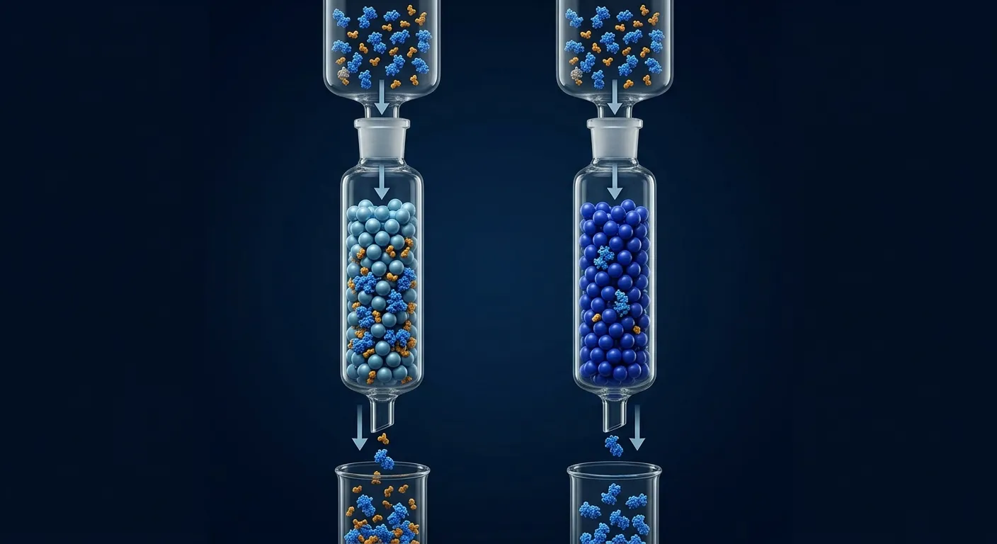
You expressed your His6-tagged protein. The lysate is loaded onto the Ni-NTA column. You elute with imidazole and... nothing comes off. Or worse: your protein is in the flowthrough, happily ignoring the nickel. The tag is there (you sequence-verified it). The column is charged (you followed the protocol). But the binding just doesn't happen.
This is one of the most common problems in recombinant protein purification, and it almost always has a simple, fixable cause.
Key Takeaways
-
The His-tag is buried or inaccessible in the folded protein—this is the #1 cause
-
Imidazole in the lysis buffer is too high, competing with the tag before binding occurs
-
The tag was cleaved by host proteases or lost during expression—check by Western blot with anti-His antibody
-
EDTA or other chelators in buffers strip the nickel from the resin
-
Wrong terminus: some proteins need the tag at the opposite end for accessibility
The Eight Causes and Fixes
Cause 1: Tag Is Buried (Most Common)
The His6 tag is short (6 residues). If it's at a terminus that folds back against the protein surface, the histidines can't reach the nickel.
Diagnosis: Protein is in the flowthrough. Western blot confirms the tag is present.
Fixes:
-
Switch terminus (N-terminal → C-terminal or vice versa)
-
Add a flexible linker (GSGSGS) between the protein and the tag
-
Use a longer tag (His8 or His10)—more histidines = higher effective affinity even if partially occluded
-
Try denaturing purification (6 M urea or 8 M GuHCl)—if the protein binds under denaturing conditions, the tag is buried in the native state
Cause 2: Too Much Imidazole in Wash/Lysis Buffer
Many protocols include 10–20 mM imidazole in the lysis buffer to reduce nonspecific binding. But for weakly binding proteins, this is enough to prevent binding entirely.
Diagnosis: Protein binds when you remove imidazole from lysis buffer.
Fix: Drop lysis buffer imidazole to 5 mM, or remove it entirely. Accept slightly dirtier eluate and clean up with a second chromatography step.
Cause 3: Tag Lost to Proteolysis
E. coli proteases can clip the terminus, removing the His-tag during expression or lysis.
Diagnosis: Anti-His Western blot is negative, but anti-protein antibody (or Coomassie) shows the protein is present.
Fixes:
-
Use protease inhibitors during lysis (PMSF + EDTA-free cocktail)
-
Express in protease-deficient strains (BL21, which lacks Lon and OmpT)
-
Shorten expression time
-
Move the tag to the other terminus (if one end is more protease-accessible)
Cause 4: EDTA or Chelators in Buffers
EDTA chelates Ni²⁺, stripping the metal from the resin. Even 1 mM EDTA will destroy Ni-NTA binding.
Diagnosis: Protein doesn't bind, AND the resin has turned white (lost its blue/green color).
Fix: Remove ALL EDTA from lysis and binding buffers. If you need a chelator for protease inhibition, use EDTA-free protease inhibitor cocktails. Recharge the resin with NiSO₄ after EDTA exposure.
Cause 5: Wrong pH
His-tag binding requires the imidazole side chain to be deprotonated (neutral). The pKa of histidine is ~6.0. Below pH 6, histidines become protonated and lose affinity for nickel.
Diagnosis: Protein binds at pH 8.0 but not at pH 6.0.
Fix: Keep binding buffer at pH 7.5–8.0. If your protein requires lower pH, consider switching to a non-histidine affinity tag (Strep-tag II, FLAG).
Cause 6: Resin Is Exhausted or Degraded
Ni-NTA resin has finite capacity (~5–40 mg/mL depending on the resin). Old resin, or resin that's been stripped by chelators or harsh cleaning, loses binding capacity.
Diagnosis: Known His-tagged control protein also doesn't bind.
Fix: Recharge with 100 mM NiSO₄ → wash with water → equilibrate. Or use fresh resin. For batch binding, use 1 mL resin per 10–30 mg expected protein.
Cause 7: Protein Is in Inclusion Bodies
If your protein is in inclusion bodies, it's in the pellet—not the supernatant you loaded onto the column.
Diagnosis: SDS-PAGE of the insoluble pellet fraction shows a strong band at the expected MW. The soluble fraction (what you loaded) has little or no target protein.
Fix: Either solubilize inclusion bodies (8 M urea, purify under denaturing conditions, refold) or address the solubility problem (lower temperature, fusion partner, different construct).
Cause 8: High Endogenous Histidine-Rich Proteins Competing
Some E. coli proteins (SlyD, ArnA, and others) have histidine-rich regions that bind Ni-NTA. At high expression, they can saturate the resin before your target binds.
Diagnosis: Eluate contains many bands, but your target is faint or absent. The flowthrough contains your protein.
Fix: Increase resin volume. Or use BL21(DE3) ΔSlyD strains. Or add 20 mM imidazole to the lysis buffer to selectively reduce contaminant binding (if your protein can handle it).
Quick Diagnostic Table
| Observation | Most Likely Cause | First Thing to Try |
|---|---|---|
| Protein in flowthrough, tag confirmed by Western | Tag buried | Switch terminus or add linker |
| Protein in flowthrough, no tag on Western | Proteolysis | Protease inhibitors, shorter expression |
| Nothing binds, resin is white | EDTA stripped the nickel | Remove EDTA, recharge resin |
| Protein binds at pH 8 but not pH 6 | Histidine protonation | Use pH 7.5–8.0 |
| Protein in pellet, not supernatant | Inclusion bodies | Solubilize or fix solubility |
| Control protein also doesn't bind | Dead resin | Fresh resin or recharge |
| Lots of contaminants, target diluted out | Resin saturated by E. coli proteins | More resin, or 20 mM imidazole wash |
The Bottom Line
| Problem | First Diagnostic | Fix |
|---|---|---|
| Tag is present but protein doesn't bind | Western blot + denaturing purification test | Switch terminus; add GS linker; use His10 |
| Tag absent on Western blot | Compare lysate fractions by Western | Add protease inhibitors; use BL21; shorten expression |
| Resin looks white/washed out | Check buffer composition for EDTA | Remove EDTA; recharge resin with NiSO₄ |
| Protein runs through at low pH | Test binding at pH 7.5–8.0 | Maintain pH ≥ 7.5 or switch to Strep-tag II |
| Protein absent from soluble fraction | SDS-PAGE of pellet vs supernatant | Address inclusion bodies (lower temp, chaperones) |
| E. coli proteins saturate the column | Run known His-tagged control | More resin + 20 mM imidazole in wash |
The one rule: His-tag purification fails for mechanical reasons, not magical ones. Systematic diagnosis—Western blot to confirm the tag, SDS-PAGE of soluble vs insoluble fractions, pH check, EDTA check—identifies the cause in under an hour.
Designing Better Constructs from the Start
Orbion's Construct Design module lets you choose tag placement (N- or C-terminal), select from a library of purification tags and fusion partners, and add flexible linkers—all informed by the protein's predicted structure. AstraPTM flags potential protease cleavage sites near the terminus, and AstraSUIT predicts expression suitability, helping you avoid the inclusion body problem before it starts.
References
-
Bornhorst JA & Bhatt S. (2000). Purification of proteins using polyhistidine affinity tags. Methods in Enzymology, 326:245-254. Link
-
Robichon C, et al. (2011). Engineering Escherichia coli BL21(DE3) derivative strains to minimize E. coli protein contamination after IMAC purification. Applied and Environmental Microbiology, 77(13):4634-4646. PMC3127699
-
Spriestersbach A, et al. (2015). Purification of His-tagged proteins. Methods in Enzymology, 559:1-15. Link