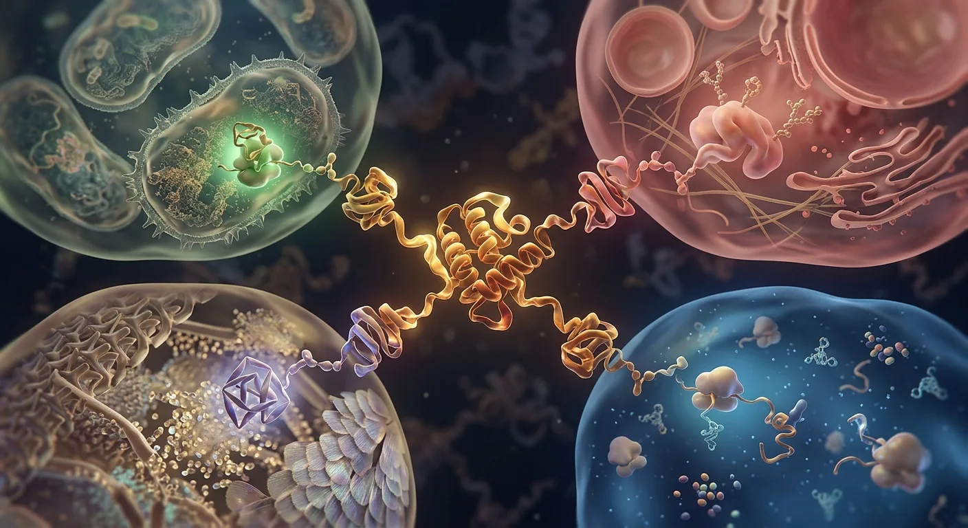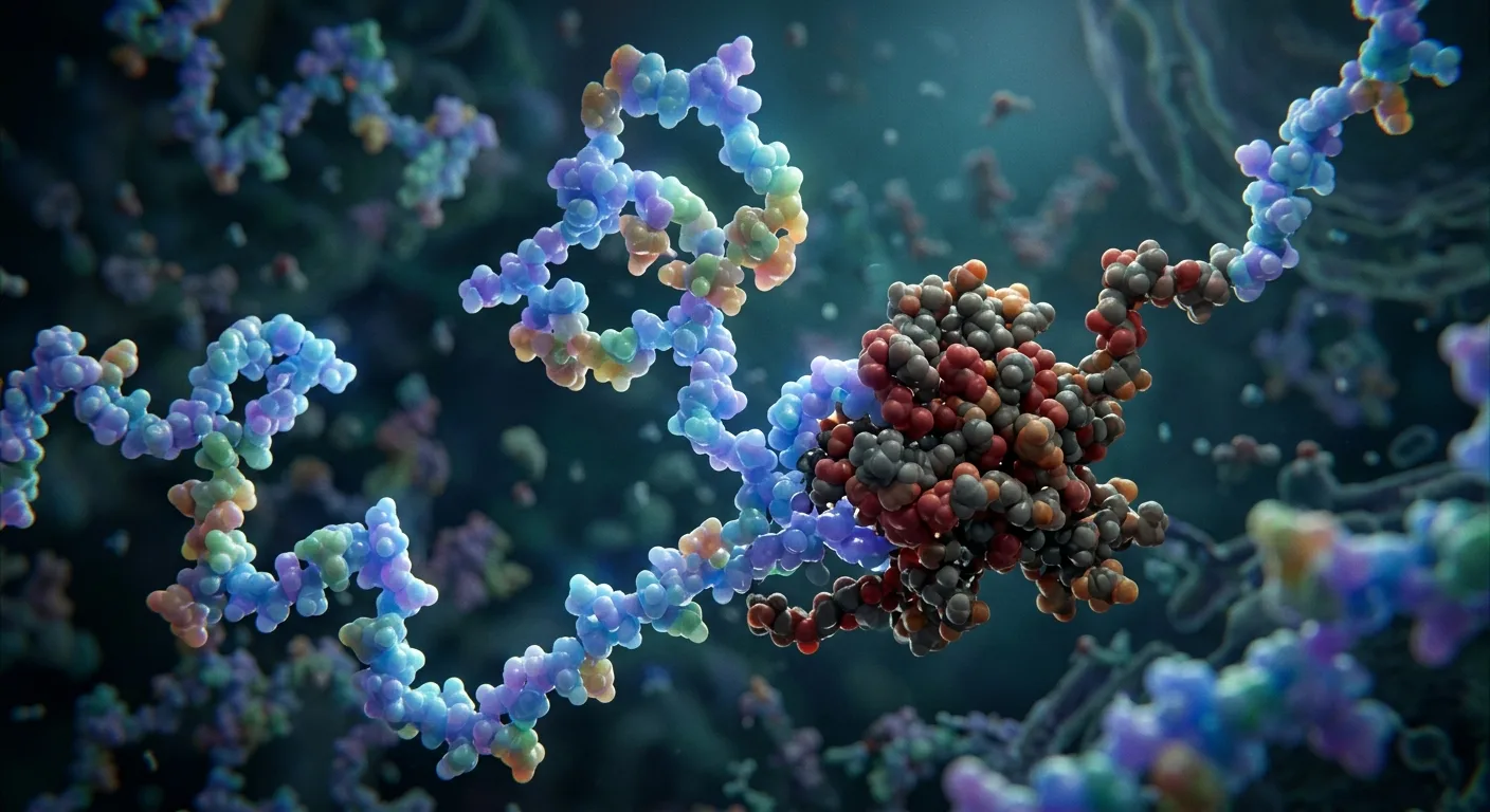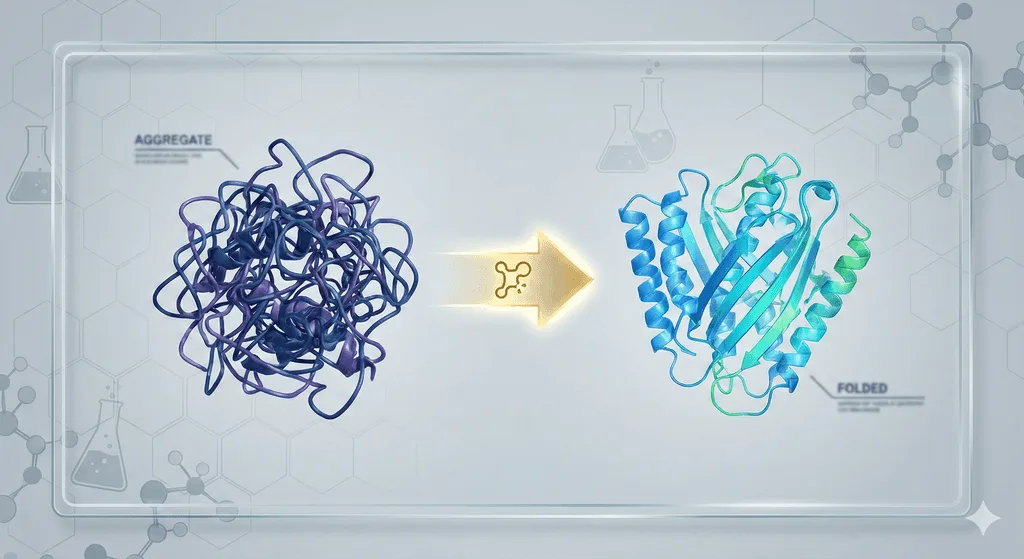
You expressed your protein. It looked perfect in gel filtration. Then you concentrated it to 5 mg/mL and found a white precipitate at the bottom of the tube. Welcome to the most frustrating problem in protein science: aggregation. It kills more projects than any other technical failure, costs labs hundreds of thousands of dollars in wasted time, and is the reason 30-40% of potential drug targets are labeled "undruggable." Here's why it happens—and how to fix it.
Key Takeaways
-
60-80% of recombinant proteins show some aggregation during expression or purification
-
Main culprits: Exposed hydrophobic patches, partial unfolding, concentration-dependent oligomerization
-
Cost of failure: $50k-100k per failed protein target (6-12 months of wasted effort)
-
Solution: Computational prediction of aggregation hotspots + rational mutagenesis increases success rates 2-5×
-
Modern tools: AI-driven prediction (Orbion, AGGRESCAN3D, CamSol) dramatically reduces failure rates
The Aggregation Problem: Why It Matters
By the Numbers
Academic research:
-
40% of proteins in structural genomics pipelines fail due to aggregation
-
Average time lost per failed target: 6-12 months
-
Opportunity cost: Cannot pursue alternative targets
Biopharmaceuticals:
-
FDA requires <2% aggregation for therapeutic antibody approval
-
High-concentration formulations (>100 mg/mL for subcutaneous injection) are aggregation-prone
-
Market impact: A rejected antibody formulation = $500M-1B loss
Industrial enzymes:
-
Aggregation during fermentation reduces yield 50-90%
-
Stability loss during storage renders products unusable
-
Economic impact: $30-50k per failed batch
The pattern: Aggregation isn't a niche problem. It's the primary technical barrier in protein production, structural biology, and therapeutics.
The Three Types of Aggregation
Not all aggregation is the same. Understanding the mechanism helps you choose the right fix.
Type 1: Co-Translational Aggregation (Inclusion Bodies)
When it happens: During protein expression in the cell (usually E. coli)
What you see: Insoluble protein pellet after cell lysis. Protein is in the insoluble fraction on SDS-PAGE.
Mechanism:
-
Protein folds faster than chaperones can assist
-
Hydrophobic regions exposed during folding stick together
-
Misfolded intermediates aggregate irreversibly
Prevalence:
-
60-70% of membrane proteins in E. coli
-
30-40% of eukaryotic proteins in E. coli
-
Less common in eukaryotic expression (yeast, insect, mammalian cells have better chaperone machinery)
Example: GPCRs in E. coli G-protein coupled receptors are 7-transmembrane proteins. When expressed in bacteria:
-
Lack of mammalian chaperones
-
No membrane insertion machinery
-
Hydrophobic transmembrane helices aggregate instantly
-
Result: 100% insoluble
Solution pathway:
-
Try lower temperature expression (16°C vs 37°C) - slower folding, more time for chaperones
-
Co-express chaperones (GroEL/ES, DnaK/J)
-
Switch to eukaryotic system (Pichia, insect cells, HEK293)
-
Add solubility tags (MBP, SUMO, GST)
Type 2: Post-Purification Aggregation (Concentration-Induced)
When it happens: During concentration, storage, or freeze-thaw
What you see:
-
Protein is initially soluble after purification
-
Concentrating above threshold (e.g., >2 mg/mL) causes precipitation
-
Cloudy solution, white precipitate, loss of activity
Mechanism:
-
Protein-protein collisions increase with concentration (C²)
-
Transient unfolding exposes aggregation-prone regions (APRs)
-
Oligomers form → grow into larger aggregates → precipitate
Prevalence:
-
~50% of antibodies show concentration-dependent aggregation
-
Therapeutic proteins requiring high concentration (>50 mg/mL) almost always face this
-
Storage-induced: Freeze-thaw cycles cause partial unfolding
Example: Therapeutic Antibody Formulation Monoclonal antibodies for subcutaneous injection need >100 mg/mL:
-
At 10 mg/mL: Stable for months
-
At 100 mg/mL: Aggregates form within days
-
At 150 mg/mL: Immediate precipitation
FDA requirement: <2% high-molecular-weight species (aggregates) at shelf life
Solution pathway:
-
Identify aggregation hotspots (hydrophobic surface patches)
-
Introduce surface mutations to reduce patch size (hydrophobic → polar)
-
Optimize buffer (pH, salt, additives like arginine or sucrose)
-
Use stabilizing excipients (polysorbate 80, trehalose)
Type 3: Conformational Aggregation (Partially Unfolded Intermediates)
When it happens: During purification, storage, or any stress condition (heat, pH shift, detergent removal)
What you see:
-
Protein loses activity even if it stays in solution
-
Oligomers form (dimers, trimers) that grow over time
-
Often irreversible
Mechanism:
-
Protein partially unfolds (loses tertiary structure but retains secondary structure)
-
Core hydrophobic residues become exposed
-
These exposed regions drive aggregation
Triggers:
-
Heat: Tm exceeded (even briefly)
-
pH extremes: Away from optimal pH
-
Detergent removal: Membrane proteins lose stabilizing detergent
-
Freeze-thaw: Ice crystal formation causes local denaturation
Example: GPCR Detergent Extraction β2-Adrenergic receptor:
-
In membrane: Stable, functional
-
After detergent extraction: Partially unfolds within minutes
-
Without ligand/stabilization: Aggregates within hours
-
Solution: Add antagonist ligand (stabilizes conformation) + thermostabilizing mutations
The Molecular Basis: Why Proteins Aggregate
At the molecular level, aggregation is driven by one principle: Hydrophobic effect.
Hydrophobic Patches: The Primary Culprit
Proteins fold to bury hydrophobic residues (Ile, Leu, Val, Phe, Trp, Met) in the core, exposing polar/charged residues (Lys, Arg, Glu, Asp, Ser, Thr) on the surface.
When aggregation occurs:
-
Protein partially unfolds (or folds incompletely)
-
Core hydrophobic residues become exposed
-
These patches stick to each other to minimize water contact
-
Oligomers form → grow → precipitate
Quantifying exposure:
-
SASA (Solvent-Accessible Surface Area): Hydrophobic SASA >500 Ų = aggregation risk
-
Hydrophobic moment: Amphipathic helices with hydrophobic face exposed aggregate easily
Aggregation-Prone Regions (APRs)
Certain sequences are intrinsically aggregation-prone, even in the folded state.
Characteristics of APRs:
-
High in hydrophobic residues (especially β-sheet forming: Ile, Val, Phe, Tyr)
-
Low charge (Lys, Arg, Glu, Asp prevent aggregation via electrostatic repulsion—charged residues create repulsive forces that keep protein molecules apart)
-
β-sheet propensity (amyloid-like aggregation)
Tools to identify APRs:
-
TANGO: Predicts β-sheet aggregation propensity
-
AGGRESCAN: Aggregation propensity from sequence
-
Zyggregator: Kinetic model of aggregation
-
Orbion: Structure + sequence analysis to identify surface-exposed APRs
Example: Amyloid-β (Alzheimer's disease) Amyloid-β peptide (40-42 amino acids):
-
Residues 17-21 (LVFFA): Highly hydrophobic, β-sheet prone
-
This region drives aggregation into amyloid fibrils
-
Mutations in this region (e.g., F19S) reduce aggregation
The Aggregation Pathway
Aggregation typically follows a stepwise mechanism:
Native ↔ Intermediate → Irreversible Aggregate
-
Native state: Properly folded, functional protein
-
Partially unfolded intermediate: Transient state with exposed hydrophobic regions (reversible)
-
Oligomers: Small clusters (dimers, trimers) that can grow
-
Irreversible aggregates: Large, insoluble precipitates or amyloid fibrils
The key is that early stages (native ↔ intermediate) are reversible—this is why optimizing buffer conditions can sometimes rescue aggregation. Once large aggregates form, the process is typically irreversible.
Type-Specific Aggregation Mechanisms
Disulfide scrambling (incorrect redox states): A major but often overlooked aggregation mechanism, especially for proteins with multiple cysteines:
-
Correct state: Native disulfide bonds (Cys A-Cys B, Cys C-Cys D)
-
Scrambled state: Non-native disulfide bonds (Cys A-Cys C, Cys B-Cys D)
-
Result: Misfolded protein with exposed hydrophobic patches → aggregation
When it happens:
-
During expression in E. coli (reducing cytoplasm, incorrect oxidation in periplasm)
-
During purification (oxidation during lysis, improper reducing conditions)
-
Storage with fluctuating redox conditions
Example: Antibody aggregation
-
IgG has 16 disulfide bonds
-
If even one forms incorrectly, the domain misfolds
-
Aggregates form through exposed hydrophobic regions
Solution:
-
Maintain consistent redox conditions (add DTT or β-mercaptoethanol)
-
For secreted proteins: Express in systems with proper oxidative folding (ER of eukaryotic cells)
-
Use oxidative refolding protocols if expressed in E. coli
Crystallization vs formulation aggregation:
These are distinct phenomena often confused:
Crystallization (desired):
-
Ordered, reversible protein-protein contacts
-
Specific lattice interactions
-
High local concentration in crystal, but soluble in mother liquor
-
Goal: Regular, repeating structure for diffraction
Formulation aggregation (problematic):
-
Disordered, irreversible protein-protein contacts
-
Random oligomers and precipitates
-
Loss of solubility and function
-
Goal: Prevent entirely
Key difference: Crystallization exploits weak, specific interactions; aggregation is driven by strong, non-specific hydrophobic collapse.
Intrinsically Disordered Proteins (IDPs): A Special Case
IDPs lack stable tertiary structure but are functional. They present unique aggregation challenges:
Why IDPs aggregate:
-
High conformational flexibility exposes aggregation-prone regions
-
Cannot be "rescued" by stabilizing mutations (no stable fold to stabilize)
-
Often phase-separate into droplets (liquid-liquid phase separation, LLPS) which can mature into aggregates
Examples:
-
α-synuclein (Parkinson's disease): Disordered monomer → β-sheet oligomers → amyloid fibrils
-
Tau (Alzheimer's): Disordered in solution, aggregates into neurofibrillary tangles
-
FUS, TDP-43 (ALS): Phase-separate into stress granules, can aggregate pathologically
Prediction challenge: Standard tools assume a folded structure. For IDPs, use specialized tools:
-
PONDR, IUPred (disorder prediction)
-
CamSol (solubility)
-
Focus on sequence-based APR prediction (TANGO, AGGRESCAN)
Predicting Aggregation: The Old Way vs The New Way
The Old Way: Trial and Error
Typical workflow (2010s):
-
Clone gene → Express in E. coli
-
Inclusion bodies → Try refolding (10+ buffer conditions)
-
Still insoluble → Try solubility tags (MBP, GST)
-
Finally soluble → Purify
-
Concentrate → Aggregates at 2 mg/mL
-
Try 20 buffer conditions, additives, pH values
-
Still aggregates → Truncate termini, try new constructs
-
Repeat for 6-12 months
Cost:
-
PhD student/postdoc time: $60k/year
-
Reagents, expression trials: $20-30k
-
Opportunity cost: Cannot pursue other targets
-
Total: $80-100k per failed protein
Success rate: ~30-40% for difficult targets (GPCRs, membrane proteins, intrinsically disordered proteins)
The New Way: Computational Prediction
Modern workflow (2024):
-
Analyze sequence for aggregation-prone regions (APRs)
-
Predict structure (AlphaFold if no experimental structure)
-
Identify hotspots: Surface-exposed hydrophobic patches
-
Design mutations: Replace aggregation-prone residues with polar/charged residues
-
Express optimized construct → Soluble, stable protein
Cost:
-
Computational analysis: Hours (free tools) to days (Orbion)
-
Optimized construct design: 1-2 weeks
-
Expression testing: 2-4 weeks
-
Total: <$5k, 4-8 weeks
Success rate improvement: 2-5× higher success rate for difficult targets (when computational design is used)
ROI: 5-10× reduction in cost, 3-5× faster
Tools for Aggregation Prediction
Sequence-Based Tools (No Structure Required)
1. AGGRESCAN
-
What it does: Scans sequence for aggregation-prone regions
-
Output: Aggregation propensity score per residue
-
Best for: Quick initial screen
-
Limitation: Doesn't account for burial in folded structure
2. TANGO
-
What it does: Predicts β-sheet aggregation (amyloid-like)
-
Output: Aggregation rate constants
-
Best for: Identifying amyloidogenic segments
-
Limitation: Focused on β-sheet aggregation (misses other types)
3. Zyggregator
-
What it does: Kinetic model of aggregation based on physicochemical properties
-
Output: Aggregation rate at different concentrations/temperatures
-
Best for: Predicting concentration dependence
-
Limitation: Empirical model, less accurate for membrane proteins
Structure-Based Tools (Requires 3D Structure)
4. AGGRESCAN3D
-
What it does: Maps aggregation propensity onto 3D structure
-
Output: Surface-exposed aggregation-prone patches
-
Best for: Identifying regions to mutate
-
Limitation: Requires accurate structure
5. CamSol (Cambridge Solubility Predictor)
-
What it does: Predicts protein solubility from structure
-
Output: Solubility score, identifies problematic residues
-
Best for: Antibody and therapeutic protein optimization
-
Limitation: Trained mostly on antibodies
6. Spatial Aggregation Propensity (SAP)
-
What it does: Analyzes clustering of hydrophobic residues on surface
-
Output: Hotspot map
-
Best for: Detecting non-obvious aggregation mechanisms
AI/ML Platforms (Structure + Sequence + Context)
7. Orbion
-
What it does:
-
Predicts aggregation hotspots from AlphaFold or experimental structure
-
Suggests specific mutations to reduce aggregation (with ΔΔG predictions)
-
Integrates with stability predictions (ensure mutations don't destabilize fold)
-
Recommends expression system based on aggregation risk
-
-
Output:
-
Aggregation risk score
-
Residue-level hotspot map
-
Mutation recommendations (e.g., "L47S reduces aggregation 80%, ΔΔG = +0.3 kcal/mol, maintains stability")
-
-
Best for: Rescue strategies for failed targets, therapeutic antibody optimization
-
Advantage: Combines multiple prediction methods + experimental validation data
How to Fix Aggregation: The Rescue Toolkit
Once you know why your protein aggregates, you can fix it. Here are the five main strategies:
Strategy 1: Mutation to Remove Aggregation Hotspots
Goal: Replace hydrophobic surface residues with polar/charged residues
Workflow:
-
Identify surface-exposed hydrophobic patches (AGGRESCAN3D, Orbion)
-
Select residues to mutate (prioritize Leu, Ile, Val, Phe, Met on surface)
-
Choose replacement residues:
-
Hydrophobic → Polar: Leu → Ser, Ile → Thr
-
Hydrophobic → Charged: Val → Glu, Phe → Asp (if structure allows)
-
-
Check that mutation doesn't destabilize fold (Orbion ΔΔG prediction, or Rosetta)
-
Make 2-3 mutants, test in parallel
Example: Antibody VH Domain Aggregation
Problem:
-
Therapeutic antibody aggregates above 50 mg/mL
-
AGGRESCAN3D identifies hotspot in CDR2 (VH residues 52-56: YISSG)
-
Hydrophobic Tyr52 and Ile53 exposed on surface
Solution:
-
Mutate Ile53 → Ser (I53S)
-
Test: I53S variant remains soluble at 120 mg/mL
-
Binding affinity: Maintained (CDR3 dominates binding)
-
Result: Developable antibody
How Orbion helps:
-
Identifies the hotspot
-
Suggests I53S mutation
-
Predicts ΔΔG = +0.2 kcal/mol (slightly stabilizing)
-
Confirms mutation is in non-critical region (binding affinity unaffected)
Strategy 2: Surface Entropy Reduction (SER)
Concept: High-entropy surface residues (Lys, Glu with flexible side chains) can promote aggregation by increasing conformational entropy. Replacing them with low-entropy residues (Ala, Ser) reduces aggregation and improves crystallization.
When to use: Proteins that aggregate during concentration or resist crystallization
How it works:
-
Identify surface Lys or Glu residues with high B-factors (flexible)
-
Mutate to Ala or Ser (smaller, less flexible)
-
Test for solubility and crystallization
Example: T4 Lysozyme
-
Mutation: K60A, E62A, K65A
-
Result: Improved crystal quality (better diffraction), reduced aggregation
Orbion:
-
Identifies high-entropy surface residues
-
Suggests SER mutations
-
Predicts impact on stability
Strategy 3: Fusion Tags & Solubility Enhancers
Goal: Add a highly soluble protein domain to mask aggregation-prone regions
Common tags:
MBP (Maltose Binding Protein) - 42 kDa
-
Mechanism: MBP is extremely soluble; fusing it to your protein increases overall solubility
-
Best for: Cytoplasmic proteins, difficult-to-express proteins
-
Drawback: Large tag, may need removal (adds purification step)
GST (Glutathione S-Transferase) - 26 kDa
-
Mechanism: Soluble, dimerizes (can help stabilize target protein)
-
Best for: Protein-protein interaction studies, pull-down assays
-
Drawback: Dimerization may not be desired
SUMO (Small Ubiquitin-like Modifier) - 12 kDa
-
Mechanism: Enhances folding, protease cleavage leaves no scar
-
Best for: Proteins requiring native N-terminus
-
Drawback: Requires specific protease (Ulp1)
NusA - 55 kDa
-
Mechanism: Bacterial chaperone, highly soluble
-
Best for: Proteins prone to inclusion bodies
-
Drawback: Very large tag
When to use:
-
Inclusion bodies in E. coli → Try MBP or NusA
-
Aggregation after purification → Try SUMO or small tags
-
Need native termini → Use cleavable tags (TEV protease site)
Example: Membrane Protein Solubilization Human aquaporin (water channel):
-
In E. coli: 100% inclusion bodies
-
With MBP fusion: 60% soluble
-
After detergent screening: Functional protein
-
Result: Structure determination possible
Strategy 4: Expression System Optimization
Principle: Different organisms have different folding machinery, chaperones, and PTM capabilities. Switching systems can solve aggregation.
Decision tree:
E. coli (bacteria):
-
Pros: Fast, cheap, high yield
-
Cons: No eukaryotic chaperones, no glycosylation, limited disulfide formation
-
Best for: Simple cytoplasmic proteins, no PTMs needed
Pichia pastoris (yeast):
-
Pros: Eukaryotic folding, some glycosylation, disulfide bonds
-
Cons: High-mannose glycans (non-human), lower yield than E. coli
-
Best for: Secreted proteins, simple glycoproteins
Sf9/Hi5 (insect cells):
-
Pros: Good folding, complex disulfides, some glycosylation
-
Cons: Minimal sialylation, expensive
-
Best for: GPCRs, complex membrane proteins
HEK293/CHO (mammalian cells):
-
Pros: Native-like folding, full glycosylation, correct PTMs
-
Cons: Slow, expensive, lower yield
-
Best for: Therapeutic antibodies, membrane proteins requiring native PTMs
Example: GPCR Expression β2-Adrenergic receptor:
-
E. coli: Inclusion bodies (100%)
-
Pichia: Partially functional (20% yield)
-
Sf9 (insect cells): Functional receptor (60% yield)
-
Choice: Insect cells (balance of yield and function)
How Orbion helps:
-
Analyzes PTM requirements (glycosylation, phosphorylation)
-
Recommends expression system
-
Example: "This protein requires N-glycosylation at Asn120 → Use insect or mammalian cells"
Strategy 5: Buffer & Formulation Optimization
Goal: Optimize solution conditions to prevent aggregation
Key variables:
1. pH
-
Principle: Proteins aggregate near their pI (isoelectric point) due to reduced charge repulsion
-
Solution: Work at pH 1-2 units away from pI
-
Tool: Calculate pI (ExPASy ProtParam), test pH 5.5, 6.5, 7.5, 8.5
2. Salt concentration
-
Principle: Moderate salt (50-150 mM NaCl) screens charge interactions, reduces aggregation
-
Too low salt: Charge-charge aggregation
-
Too high salt: Salting-out effect (protein precipitates)
-
Optimal: 100-150 mM NaCl for most proteins
3. Additives
-
Arginine (50-500 mM): Suppresses aggregation (mechanism unclear, likely disrupts hydrophobic interactions)
-
Glycerol (5-20%): Stabilizes native state, reduces unfolding
-
Sucrose/Trehalose (5-10%): Osmolytes, protect against freeze-thaw
-
Detergents (membrane proteins): Maintain solubility (DDM, LMNG, amphipols)
4. Temperature
-
Cold storage (4°C): Slows aggregation kinetics
-
Flash-freezing: Rapid cooling minimizes ice crystal damage
-
Avoid freeze-thaw: Each cycle causes partial unfolding
Example: Antibody Formulation Optimization Monoclonal antibody aggregating at >100 mg/mL:
-
Initial buffer: PBS (pH 7.4, 150 mM NaCl)
-
Aggregates at 120 mg/mL
Optimization screen:
-
pH: Test 5.5, 6.5, 7.5, 8.5 → Best at pH 6.0
-
Additives: Add 10 mM arginine + 5% sucrose
-
Result: Stable at 150 mg/mL for 6 months
Case Study: Rescuing an Aggregation-Prone Therapeutic Antibody
The Challenge
Target: Therapeutic monoclonal antibody (IgG1) for cancer Problem: Aggregates above 50 mg/mL (subcutaneous formulation requires >100 mg/mL) Timeline: 8 months in, Phase I trials delayed
The Investigation
Step 1: Identify aggregation mechanism
-
DLS (dynamic light scattering): Oligomers form at >30 mg/mL
-
SEC-MALS: High-molecular-weight species (dimers, trimers)
-
Conclusion: Concentration-dependent, reversible aggregation
Step 2: Computational prediction
-
Run AGGRESCAN3D on antibody structure
-
Hotspot identified: VH CDR2 (residues 52-56)
-
Hydrophobic patch (Tyr52, Ile53, Phe55) exposed on surface
Step 3: Rational mutagenesis
-
Design mutations:
-
I53S (Ile → Ser)
-
F55T (Phe → Thr)
-
Double mutant: I53S/F55T
-
-
Predict ΔΔG (stability): Orbion predicts +0.3 kcal/mol (slightly stabilizing)
The Solution
Step 4: Experimental validation
-
Express I53S, F55T, and I53S/F55T variants
-
Test solubility at 100, 150, 200 mg/mL
Results:
-
Wild-type: Aggregates at 60 mg/mL
-
I53S: Soluble to 130 mg/mL
-
F55T: Soluble to 110 mg/mL
-
I53S/F55T: Soluble to 180 mg/mL ✓
Step 5: Functional validation
-
Binding affinity (SPR): I53S/F55T binds with same Kd as wild-type
-
ADCC activity: Maintained
-
Pharmacokinetics (mouse model): Half-life unchanged
Outcome:
-
Developable antibody formulation (150 mg/mL)
-
Phase I trials resumed
-
Time saved: 12 months (avoided complete redesign)
The Economics of Aggregation Prevention
Cost of Failure
Academic target:
-
1 year of postdoc time: $60k
-
Reagents: $20k
-
Opportunity cost: Missed publications, delayed graduation
-
Total: $80k
Therapeutic antibody:
-
Discovery program: $10-20M
-
If aggregation detected late (Phase I): $50-100M wasted
-
Delay to market: 1-2 years = $500M-1B in lost revenue
ROI of Computational Prediction
Upfront investment:
-
Orbion subscription: $X/month
-
Computational time: 1-2 days
Savings:
-
Avoid 6-12 months of trial-and-error
-
Test 2-3 designed mutants instead of 50 random constructs
-
Increase success rate from 30% → 70%
ROI: 10-20× return on investment
Practical Checklist: Preventing Aggregation
Before Expression
-
[ ] Run aggregation prediction (AGGRESCAN, Orbion)
-
[ ] Check pI (avoid working near pI)
-
[ ] Identify surface hydrophobic patches
-
[ ] Design 2-3 mutants if hotspots found
-
[ ] Choose expression system based on PTM needs
During Expression
-
[ ] Small-scale test (don't commit to 10L culture immediately)
-
[ ] Check soluble vs insoluble fractions (SDS-PAGE)
-
[ ] If insoluble: Try 16°C expression, co-express chaperones, or add fusion tag
-
[ ] If soluble but low yield: Optimize induction conditions (IPTG concentration, time)
During Purification
-
[ ] Keep protein cold (4°C throughout)
-
[ ] Add stabilizers to buffer (glycerol 10%, DTT if cysteines present)
-
[ ] Avoid high protein concentrations early (dilute after elution)
-
[ ] Run SEC (size-exclusion chromatography) to remove aggregates
During Storage
-
[ ] Aliquot and flash-freeze (minimize freeze-thaw cycles)
-
[ ] Add cryoprotectants (10% glycerol or 5% trehalose)
-
[ ] Store at -80°C (more stable than -20°C)
-
[ ] For long-term: Lyophilize (freeze-dry)
If It Still Aggregates
-
[ ] Try fusion tag (MBP, SUMO)
-
[ ] Switch expression system (E. coli → yeast → insect → mammalian)
-
[ ] Revisit construct design (truncate disordered regions)
-
[ ] Screen formulation buffers (pH, salt, additives)
The Future: AI-Driven Aggregation Rescue
Machine learning models trained on millions of protein sequences and structures are making aggregation prediction more accurate.
Emerging Tools
1. AlphaFold confidence scores as aggregation predictors
-
Low pLDDT regions (<70) often correlate with aggregation-prone loops
-
Not explicit aggregation prediction, but useful heuristic
2. Generative models for solubility optimization
-
Tools like ProteinMPNN can redesign surface to maximize solubility
-
Generate variants with same function, improved expression
3. Deep learning scoring functions
-
Train on experimental aggregation data
-
Predict aggregation rate at different concentrations
4. Integration platforms (like Orbion)
-
Combine structure, sequence, PTM prediction, stability
-
Suggest complete rescue strategy (mutations + expression system + formulation)
The Bottom Line
Protein aggregation is not random bad luck. It is a predictable, solvable engineering problem.
The old paradigm: Express → Hope it works → Troubleshoot for months
The new paradigm: Predict → Design → Express optimized construct → Success
With modern computational tools, you can:
-
Predict aggregation before expressing anything
-
Design rescue mutations to remove hotspots
-
Choose the right expression system based on PTM and folding needs
-
Optimize formulation computationally before experimental screening
The payoff:
-
2-5× improvement in success rates
-
3-5× faster timelines
-
10× cost reduction
The difference between a "difficult protein" and a "successful structure" is often just 1-2 well-placed mutations identified by prediction tools like Orbion.
Aggregation used to be the graveyard of protein projects. Today, it's dramatically reduced—if you use the right tools. While not every aggregation problem can be solved computationally, the success rate has improved dramatically, transforming once-intractable targets into achievable goals.
Ready to Rescue Your Aggregating Protein?
If you have a protein that aggregates during expression, purification, or storage, Orbion can help identify why and suggest how to fix it.
Orbion provides:
-
Aggregation hotspot prediction from structure or sequence
-
Specific mutation recommendations (with ΔΔG stability predictions)
-
Expression system selection based on PTM requirements
-
Formulation optimization guidance