Your His-tagged protein came off the Ni-NTA looking clean, mostly. There is your band at the expected mass, and then there is that other one: a fat, stubborn contaminant sitting around 60 kDa that no amount of imidazole washing seems to shift. You did not clone a 60 kDa passenger. You did not add one. But it co-eluted with your protein, it scales with your protein, and on a good prep it can be 10 to 30% of the total stained material. That band is almost certainly GroEL, the E. coli chaperonin, and its presence is not just a purity nuisance. It is a message about how your protein is folding.
Chaperone co-purification is one of the most misread results in recombinant protein production. People treat it as a chromatography problem to wash away. It is really a folding problem wearing a chromatography costume. This article covers who these contaminants are, the two distinct reasons they ride along through IMAC, how to confirm the diagnosis, how to remove them, and, most importantly, what you should change upstream so they stop showing up.
This is not the tag-removal problem (protease will not cut) or the endotoxin problem (LPS ruins your cell assay). Those are separate posts. This is about the E. coli folding machinery hitching a ride on your prep.
Key Takeaways
- The ~60 kDa band is usually GroEL. The E. coli chaperonin subunit is 57 kDa and assembles into an 800 kDa tetradecamer; its co-chaperonin GroES is 10 kDa, and the Hsp70 chaperone DnaK runs at 70 kDa. These three account for the large majority of chaperone contamination in IMAC preps.
- GroEL is a normal client of hundreds of E. coli proteins. Proteome-wide analysis identified roughly 250 GroEL-interacting proteins, of which about 85 are obligate clients that essentially require the chaperonin to fold (Kerner et al., 2005). Your recombinant protein can join that list.
- Two mechanisms drive co-purification, and they need different fixes. Chaperones either bind the Ni resin directly through native histidine clusters, or they hitchhike bound to your misfolded protein. Roughly 40% of common E. coli IMAC contaminants show strong intrinsic metal affinity (Bolanos-Garcia & Davies, 2006).
- An ATP plus Mg wash is both a cure and a diagnostic. Chaperones capture clients in an ATP-independent step, so they persist through standard washes; adding 5 mM ATP with 10 mM MgCl2 on-column triggers client release and strips them off. If the band disappears with Mg-ATP, it was a chaperone.
- Chaperones on your prep mean your protein is folding poorly. They bind exposed hydrophobic surface, the signature of folding intermediates, aggregation-prone regions, and misfolded chains. Removing the band does not fix the underlying instability.
- The durable fix is upstream. Lower induction temperature, slower induction, solubility fusions, and construct redesign reduce the exposed hydrophobicity that recruits chaperones in the first place.
Introduction
Immobilized metal affinity chromatography (IMAC) works because a run of histidines binds nickel or cobalt more tightly than almost anything else in an E. coli lysate. "Almost" is the operative word. E. coli makes its own proteins with surface-exposed histidine clusters, and it makes molecular chaperones whose entire job is to grab onto partially folded client proteins. Put those two facts together on a Ni-NTA column and you get co-purification.
Recombinant expression makes this worse, not better. When you drive a heterologous protein to high levels in E. coli, a substantial fraction of it does not reach the native state cleanly. Overexpression floods the folding capacity of the cell, and the chaperone network responds by binding the backlog. Around 30% of recombinant proteins fail to express in soluble, usable form in E. coli (Baneyx & Mujacic, 2004), and much of the soluble fraction that does appear passes through chaperone-bound intermediates on its way to folding, or gets stuck there. Those chaperone-client complexes are stable enough to survive lysis, loading, and washing.
The result is predictable. You see GroEL at 60 kDa, sometimes GroES at 10 kDa near the dye front, and often DnaK at 70 kDa. The interesting question is not "how do I get rid of it" but "why is it there, and what is it telling me about my protein."
The Usual Suspects: GroEL, GroES, and DnaK
Three chaperones account for most of what co-purifies. Knowing their exact masses lets you read a gel at a glance.
| Chaperone | Subunit mass | Assembly | Role |
|---|---|---|---|
| GroEL (Hsp60 / chaperonin) | 57 kDa | 14-mer, ~800 kDa double ring | Encloses client in a folding chamber; ATP-driven |
| GroES (Hsp10 / co-chaperonin) | 10 kDa | 7-mer, ~70 kDa | Caps the GroEL ring, gates the chamber |
| DnaK (Hsp70) | 70 kDa | Monomer (cycles with DnaJ, GrpE) | Binds extended hydrophobic segments on nascent and stressed chains |
GroEL is the one you see most. Its 57 kDa subunit runs right in the middle of a typical gel, and because the ring is so abundant under stress it stains heavily. GroES is easy to miss because it runs low, near small tags and dye, but it often travels with GroEL. DnaK at 70 kDa is the second most common band and is the dominant contaminant of fusion proteins whose linker regions present DnaK-binding motifs (Rial & Ceccarelli, 2002).
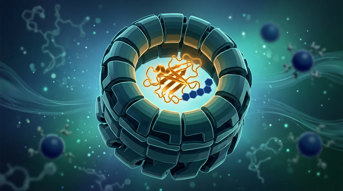
For mechanism, GroEL and DnaK work differently. DnaK (Hsp70) grabs short, extended, hydrophobic stretches (roughly 5 to 7 residues enriched in Leu, Ile, Val, Phe flanked by basic residues) as chains emerge or unfold. GroEL takes larger, compact but non-native folding intermediates and encloses them in its chamber for an ATP-gated folding attempt (Hartl, Bracher & Hayer-Hartl, 2011). Both bind exposed hydrophobicity. Both hold on until ATP tells them to let go. That single shared property, ATP-triggered release, is the key to both diagnosing and removing them.
Why Chaperones Bind Your Protein in the First Place
Chaperones do not recognize your protein by sequence identity. They recognize the physical signature of an unfinished fold: hydrophobic residues that should be buried in the core but are instead exposed to solvent.
A correctly folded, stable protein buries its greasy residues. A folding intermediate, a misfolded chain, or an aggregation-prone construct leaves patches of that hydrophobic surface hanging out. To GroEL and DnaK, that surface looks exactly like a client that needs help. So they bind. The more of your protein that lingers in a partially folded state, the more chaperone it recruits.
This is why chaperone load correlates with folding difficulty. In the E. coli proteome, the obligate GroEL clients are enriched for proteins with complex topologies (notably TIM-barrel and other domains that fold slowly through aggregation-prone intermediates), and these clients are, on average, larger and more aggregation-prone than the general proteome (Kerner et al., 2005). When you overexpress a difficult heterologous protein, you are manufacturing exactly the substrate the chaperone system evolved to grab.
The practical consequence: the amount of GroEL on your gel is a rough readout of how much of your protein failed to fold cleanly. A faint trace is normal. A heavy 60 kDa band that scales with your target is a red flag.
Why They Survive IMAC: Two Independent Mechanisms
Here is the part that trips people up. There are two completely different reasons a chaperone ends up in your elution, and they call for different fixes.
Mechanism 1: The chaperone binds the resin directly
E. coli chaperones and several other abundant proteins carry surface-exposed histidine clusters that bind Ni-NTA and Co-NTA on their own, with no help from your protein. In a systematic analysis of native E. coli proteins that contaminate IMAC preps, about 40% showed strong affinity for metal-chelating resin, 47% moderate, and 13% weak (Bolanos-Garcia & Davies, 2006). GroEL, DnaK, and the peptidyl-prolyl isomerase SlyD are recurring offenders. SlyD in particular is histidine-rich and clings tenaciously even at high imidazole.
If this is your mechanism, the contaminant is present whether or not your protein misfolds. It is an intrinsic property of the E. coli proteome meeting a nickel column.
Mechanism 2: The chaperone hitchhikes on your misfolded protein
The second route is the one that carries diagnostic weight. Your His-tagged protein binds the resin through its tag, as intended. But your protein is also, in part, a folding client, so it arrives at the column already bound to GroEL or DnaK. The chaperone is not on the resin because of its own metal affinity. It is there because it is physically clamped onto your protein, and your protein is bolted to the nickel.
Crucially, this capture is ATP-independent. Chaperones bind their clients tightly in the nucleotide-free or ADP state. Release requires ATP. Your wash buffers contain no ATP, so the complex stays intact through loading, washing, and elution. The chaperone rides your protein straight into the tube.
Distinguishing the two mechanisms matters because Mechanism 1 is fixed by chromatography alone, while Mechanism 2 is fixed by biology: if you improve folding, the hitchhiker disappears.
How to Confirm It Really Is a Chaperone
Before you spend a week chasing a contaminant, confirm what it is. A ~60 kDa band is not automatically GroEL. It could be a degradation product, a dimer of a 30 kDa target, or a co-purifying host enzyme.
Signatures that point to GroEL:
- Mass. GroEL runs at 57 to 60 kDa, sharp and reproducible. GroES at ~10 kDa near the dye front is a strong corroborating sign because the two travel together.
- It scales with expression stress, not with your protein's yield. Heavier on a hot induction, lighter on a cool one.
- It resists imidazole washes that remove weaker contaminants, because it is either intrinsically metal-bound or clamped onto your target.
Definitive tests:
- Mass spectrometry (the gold standard). Excise the band, trypsin digest, and run peptide mass fingerprinting or LC-MS/MS. GroEL, GroES, DnaK, and SlyD all have well-characterized peptide signatures and will be identified unambiguously. This is the only way to be certain.
- The Mg-ATP release test (fast and cheap). Split your bound resin. Wash one half normally; wash the other with buffer containing 5 mM ATP and 10 to 20 mM MgCl2 (add ~50 to 150 mM KCl for DnaK). Incubate 10 to 30 minutes at room temperature, then elute both. If the 60 kDa band drops sharply in the ATP-treated half, it was a chaperone caught in the act of holding a client (Mechanism 2), because ATP triggered release. If the band is unchanged, you are looking at direct resin binding (Mechanism 1) or a non-chaperone contaminant.
- Western blot with anti-GroEL or anti-DnaK antibodies (commercially available) confirms identity without mass spec if you already suspect a specific chaperone.
The Mg-ATP test does double duty: it tells you what the band is and which removal strategy will work.
What the Chaperone Is Actually Telling You
This is the section most troubleshooting guides skip, and it is the most important one.
If your contaminant is Mechanism 2 (a hitchhiker on your protein), then GroEL on your gel is a direct, physical report that a measurable fraction of your protein is not natively folded. The chaperone is bound to exposed hydrophobic surface that a properly folded protein would have buried. You can wash the chaperone away, but the protein it was holding is still the same poorly folded, aggregation-prone molecule. You have cleaned up the gel, not the protein.
This has real downstream consequences. Protein that co-purified with chaperones is more likely to:
- Aggregate on concentration. The exposed hydrophobic surface that recruited GroEL is the same surface that drives aggregation once you remove the chaperone and push concentration up.
- Show heterogeneous behavior in SEC. A shoulder or a void-volume peak often traces back to the chaperone-prone fraction.
- Underperform in functional assays. Misfolded protein is inactive protein.
- Fail to crystallize. Conformational heterogeneity is the enemy of a well-diffracting lattice.
So the honest reading of a heavy chaperone band is: a chromatographic wash is a bandage; the fix is to fold more of your protein correctly. That reframing is what separates a prep you salvage once from a construct you actually improve.
How to Remove Chaperones From the Prep
When you do need to clean up an existing prep, match the method to the mechanism.
On-column Mg-ATP wash (best first move for Mechanism 2)
Because chaperone-client capture is ATP-dependent for release, feeding ATP to the loaded column pops the client loose. This is the single most effective, gentlest chaperone-stripping step.
Protocol:
- Load lysate onto Ni-NTA as usual.
- Wash with 10 to 20 CV of buffer containing 5 to 10 mM ATP, 10 to 20 mM MgCl2, and 50 to 150 mM KCl, at room temperature.
- Optionally pause the flow for 10 to 15 minutes to let release cycles complete.
- Follow with 5 CV of standard wash to clear released chaperone and nucleotide.
- Elute your protein.
Magnesium is required (ATP hydrolysis needs Mg2+); potassium markedly stimulates DnaK. Skip either and the wash underperforms.
Denaturing IMAC (nuclear option for Mechanism 1 or heavy contamination)
If the chaperone binds the resin intrinsically or is bound to protein you plan to refold anyway, wash and elute under denaturing conditions. Loading in 6 M guanidine-HCl or 8 M urea unfolds everything; native chaperone-client complexes cannot survive, and intrinsic metal binding through folded His clusters is disrupted. Your His-tag still binds nickel because the tag itself does not need to be folded. You then refold on-column or after elution. This costs you the convenience of native purification and adds a refolding step, so reserve it for cases where you will refold regardless.
High salt and detergent washes
A wash with 0.5 to 1 M NaCl disrupts electrostatic components of chaperone-client and chaperone-resin interactions and shifts some contaminants. Low levels of non-ionic detergent (0.1% Triton X-100) can dislodge hydrophobically bound chaperone. These are partial measures: helpful as add-ons, rarely sufficient alone for a heavy GroEL band.
Orthogonal polishing steps
Because GroEL assembles into an 800 kDa particle, size-exclusion chromatography separates it cleanly from most monomeric targets: the chaperonin elutes far earlier. Ion exchange also resolves chaperones from many targets on charge. If chaperone contamination survives your affinity step, a well-chosen polishing step usually finishes the job, though at the cost of yield and time.
Method comparison
| Method | Targets | Effectiveness | Cost / Downside |
|---|---|---|---|
| On-column Mg-ATP + K wash | Hitchhiking chaperones (Mech. 2) | High for GroEL/DnaK complexes | Adds ATP/Mg/K to buffer; minimal protein loss |
| Denaturing IMAC | Both mechanisms | Very high | Requires refolding; not for fold-sensitive proteins |
| High salt / detergent wash | Partial, both | Moderate | Simple add-on; rarely sufficient alone |
| Size exclusion | GroEL (800 kDa particle) | High | Yield loss, extra step, dilution |
| Ion exchange | Charge-separable chaperones | High | Method development per protein |
Removing Chaperones at the Source
Every method above is downstream cleanup. The durable fix is to produce less chaperone-bound protein to begin with. All of these reduce the exposed hydrophobicity that recruits GroEL and DnaK.
- Lower the induction temperature. Expressing at 16 to 20°C instead of 37°C slows synthesis, gives chains more time to fold before they aggregate, and is the single most reliable lever for reducing misfolding (Rosano & Ceccarelli, 2014). Less misfolded protein means less chaperone recruitment.
- Induce slowly and lightly. Lower IPTG (0.1 to 0.25 mM) and induction at lower cell density reduce the synthesis burst that overwhelms folding capacity.
- Add a solubility fusion. MBP, SUMO, NusA, TrxA, and GB1 improve folding of the passenger and reduce the exposed hydrophobic surface. Caution: a fusion can shift the problem rather than solve it. Poorly chosen linker regions actively recruit DnaK, which is a documented cause of Hsp70 contamination in fusion protein preps (Rial & Ceccarelli, 2002). Choose the fusion and linker deliberately.
- Co-express chaperones, carefully. Deliberately over-supplying GroEL/GroES or DnaK/DnaJ/GrpE can improve folding yield, but it also raises the free chaperone pool, which can increase co-purification if folding is still incomplete. This is a tuning exercise, not a default.
- Use engineered host strains. BL21(DE3) derivatives have been engineered to remove or reduce the most common IMAC contaminants (including tagged or mutated versions of the usual histidine-rich offenders), cutting native protein carryover substantially (Robichon et al., 2011).
- Use a dedicated cleanser step. A GST-tagged DnaK-substrate "cleanser" protein can be added to sequester contaminant Hsp70 and then removed on glutathione resin, a targeted way to strip DnaK without touching your target (Morales, Parcerisa & Ceccarelli, 2019).
- Redesign the construct. If a specific region drives misfolding, trimming a disordered terminus, adjusting domain boundaries, or removing an aggregation-prone segment attacks the root cause.
The theme is consistent: chaperones bind unfinished folds, so anything that improves folding reduces the contaminant.
Case Study: The 60 kDa Band That Would Not Wash Off
Problem. A group expressed a 42 kDa cytoplasmic enzyme in BL21(DE3) with an N-terminal His6 tag, induced at 37°C. IMAC gave the target plus a heavy contaminant at 60 kDa, roughly 25% of stained material. Increasing imidazole in the wash from 20 to 50 mM barely touched it, and pushing higher started stripping the target. SEC showed a partial void-volume peak, and the protein aggregated when concentrated past 3 mg/mL.
Diagnosis. They split the loaded resin and ran the Mg-ATP release test: 5 mM ATP, 15 mM MgCl2, 100 mM KCl, 20 minutes at room temperature. In the ATP-washed fraction the 60 kDa band dropped by roughly 80%. That confirmed a hitchhiking chaperone (Mechanism 2), and mass spec on the excised band returned GroEL. The void-volume SEC peak and the concentration-dependent aggregation pointed to the same conclusion: a large fraction of the enzyme was folding through, and getting stuck in, GroEL-bound intermediates.
Solution. Rather than only bolt on the ATP wash, they treated the chaperone as a folding signal. They dropped induction to 18°C overnight with 0.1 mM IPTG, and added an N-terminal MBP fusion with a deliberately chosen non-DnaK-recruiting linker. The on-column Mg-ATP wash stayed in as insurance.
Outcome. GroEL fell from ~25% of stained material to a faint trace. The void-volume SEC peak collapsed into a single monodisperse peak. Concentration-limited aggregation resolved: the protein now went to 12 mg/mL without precipitation, and specific activity rose because more of the prep was natively folded. The chaperone band had been a symptom; fixing the fold fixed the symptom and the protein.
Practical Tools: End-to-End Decision Tree
Use this from the first suspicious gel to a clean, correctly folded prep.
START: ~60 kDa (or ~70 kDa) contaminant in your IMAC elution
│
├── STEP 1: IDENTIFY THE BAND
│   ├── Mass ~57-60 kDa? → suspect GroEL
│   ├── Mass ~70 kDa?   → suspect DnaK
│   ├── Faint ~10 kDa companion near dye front? → GroES present → confirms GroEL
│   └── Confirm by: mass spec (definitive) OR anti-GroEL/anti-DnaK western
│         └── Not a chaperone (e.g., dimer, degradation, host enzyme)?
│               → different problem; stop here
│
├── STEP 2: DETERMINE THE MECHANISM (Mg-ATP release test)
│   Split loaded resin. Wash half normally; wash half with
│   5-10 mM ATP + 10-20 mM MgCl2 + 50-150 mM KCl, 10-30 min, RT.
│   │
│   ├── Band DROPS with Mg-ATP → MECHANISM 2 (hitchhiker on your protein)
│   │     → Your protein is folding poorly. Go to STEP 3A and STEP 4.
│   │
│   └── Band UNCHANGED → MECHANISM 1 (chaperone binds resin directly)
│         → Chromatography fix only. Go to STEP 3B.
│
├── STEP 3A: STRIP HITCHHIKERS (Mechanism 2)
│   ├── Add on-column Mg-ATP + K wash to your standard protocol
│   ├── Still heavy? Add SEC (GroEL = 800 kDa, elutes early) or ion exchange
│   └── ALWAYS proceed to STEP 4 (fix the fold)
│
├── STEP 3B: STRIP DIRECT BINDERS (Mechanism 1)
│   ├── Add a stringent imidazole wash step (optimize, do not over-strip target)
│   ├── High salt (0.5-1 M NaCl) and/or 0.1% Triton X-100 wash
│   ├── Consider engineered contaminant-minimized host strain for next prep
│   ├── SlyD suspected (histidine-rich, tenacious)? → EDTA-free stringent wash / strain fix
│   └── Last resort: denaturing IMAC + refold (only if fold-tolerant)
│
└── STEP 4: FIX THE FOLD AT THE SOURCE (do this whenever Mechanism 2)
    ├── Lower induction: 16-20°C, 0.1-0.25 mM IPTG, induce at lower OD
    ├── Add a solubility fusion (MBP/SUMO/NusA/TrxA/GB1)
    │     └── Choose linker deliberately (avoid DnaK-recruiting motifs)
    ├── Consider tuned chaperone co-expression (GroEL/ES or DnaK/DnaJ/GrpE)
    ├── Redesign construct: trim disordered termini, fix domain boundaries,
    │     remove aggregation-prone segments
    └── Re-express and re-run STEP 1 to confirm the band is gone
Pre-expression checklist (prevention)
- Predicted disordered regions and aggregation-prone segments mapped before cloning
- Construct boundaries chosen to avoid exposed hydrophobic termini
- Solubility fusion selected if the target is high-risk; linker screened for DnaK motifs
- Expression host chosen with contaminant load in mind
- Induction plan set to cool, slow conditions for a difficult target
- Mg-ATP wash buffer components on hand (ATP, MgCl2, KCl) before the first prep
Economic Analysis
Chaperone contamination is cheap to diagnose and expensive to ignore.
The cost of ignoring it (treating the band as pure enough):
| Stage | Time lost | Why |
|---|---|---|
| Downstream assays on impure protein | 1-2 weeks | Chaperone-bound fraction is misfolded and inactive; results are noisy or wrong |
| Aggregation on concentration | 1 week | Prep crashes; repeat from expression |
| Failed crystallization trials | 4-8 weeks | Conformational heterogeneity kills diffraction |
| Repeat prep after realizing the problem | 2-3 weeks | Back to expression |
The cost of diagnosing and fixing it:
| Stage | Time / cost | Notes |
|---|---|---|
| Mg-ATP release test | ~1 hour, a few dollars of ATP | Identifies mechanism immediately |
| Mass spec confirmation | 1-2 days, modest core-facility fee | Definitive band ID |
| Add on-column Mg-ATP wash | +30 min per prep | Negligible protein loss |
| Re-express cooler / with fusion | 1 week | Fixes the root cause |
The asymmetry is stark. A one-hour test and a one-week re-expression prevent a multi-week detour through failed assays and crystallization. The most expensive mistake is not the chaperone. It is spending months on a protein that was never properly folded, and never knowing that the 60 kDa band told you so on day one.
The Bottom Line
A chaperone band is a diagnosis, not just a contaminant. GroEL at 60 kDa, GroES at 10 kDa, and DnaK at 70 kDa co-purify because they bind the exposed hydrophobic surface of proteins that are not fully folded, and they hold on until ATP releases them, which standard washes never provide.
The workflow is short. Identify the band (mass spec, or the ~60 kDa plus ~10 kDa GroEL/GroES signature). Determine the mechanism with a Mg-ATP release test: if the band drops with ATP, it was riding your misfolded protein; if it does not, it is binding the resin on its own. For direct binders, clean up with stringent washes, orthogonal polishing, or an engineered strain. For hitchhikers, strip them with an on-column Mg-ATP plus potassium wash, and then, this is the part that matters, fix the fold upstream with cooler induction, a well-chosen solubility fusion, or construct redesign.
The mistake to avoid is treating a heavy chaperone band as a purity problem you can wash away. You can clean the gel and still be left with the same aggregation-prone, partially folded protein. The chaperone was pointing at the real issue. Listen to it.
Predict the Fold Problem Before You Express It
Chaperone co-purification is a folding red flag that shows up after weeks of cloning and expression. The cheaper move is to see the risk in the sequence before you order an oligo.
That is what Orbion's Characterization module is built for. AstraUNFOLD predicts per-residue disorder and per-residue amyloid propensity directly from sequence, flagging the exposed, aggregation-prone regions that recruit GroEL and DnaK, so you know which proteins are chaperone magnets before you express them. AstraSUIT informs expression host and experimental suitability, helping you decide whether E. coli is even the right system or whether the folding burden points elsewhere.
When a target looks risky, the Design tab lets you engineer around it. Add a solubility fusion from the component library (MBP, SUMO, NusA, TrxA, and Trigger Factor are all included), adjust construct boundaries, and score each construct on solubility, disorder, and aggregation against the wild-type reference. You choose the construct least likely to misfold, and therefore least likely to drag chaperones through your Ni-NTA column, before the first prep.
Stop reading chaperone bands after the fact. Predict the folding problem, design around it, and get a cleaner prep the first time. Upload your sequence at orbion.life.
References
-
Kerner MJ, Naylor DJ, Ishihama Y, Maier T, Chang HC, Stines AP, Georgopoulos C, Frishman D, Hayer-Hartl M, Mann M, Hartl FU. (2005). Proteome-wide analysis of chaperonin-dependent protein folding in Escherichia coli. Cell, 122(2):209-220. https://doi.org/10.1016/j.cell.2005.05.028
-
Hartl FU, Bracher A, Hayer-Hartl M. (2011). Molecular chaperones in protein folding and proteostasis. Nature, 475(7356):324-332. https://doi.org/10.1038/nature10317
-
Xu Z, Horwich AL, Sigler PB. (1997). The crystal structure of the asymmetric GroEL-GroES-(ADP)7 chaperonin complex. Nature, 388(6644):741-750. https://doi.org/10.1038/41944
-
Bolanos-Garcia VM, Davies OR. (2006). Structural analysis and classification of native proteins from E. coli commonly co-purified by immobilised metal affinity chromatography. Biochimica et Biophysica Acta (BBA) - General Subjects, 1760(9):1304-1313. https://doi.org/10.1016/j.bbagen.2006.03.027
-
Rial DV, Ceccarelli EA. (2002). Removal of DnaK contamination during fusion protein purifications. Protein Expression and Purification, 25(3):503-507. https://doi.org/10.1016/S1046-5928(02)00024-4
-
Robichon C, Luo J, Causey TB, Benner JS, Samuelson JC. (2011). Engineering Escherichia coli BL21(DE3) derivative strains to minimize E. coli protein contamination after purification by immobilized metal affinity chromatography. Applied and Environmental Microbiology, 77(13):4634-4646. https://doi.org/10.1128/AEM.00119-11
-
Morales ES, Parcerisa IL, Ceccarelli EA. (2019). A novel method for removing contaminant Hsp70 molecular chaperones from recombinant proteins. Protein Science, 28(4):800-807. https://doi.org/10.1002/pro.3574
-
Baneyx F, Mujacic M. (2004). Recombinant protein folding and misfolding in Escherichia coli. Nature Biotechnology, 22(11):1399-1408. https://doi.org/10.1038/nbt1029
-
Rosano GL, Ceccarelli EA. (2014). Recombinant protein expression in Escherichia coli: advances and challenges. Frontiers in Microbiology, 5:172. https://doi.org/10.3389/fmicb.2014.00172