Your AlphaFold model is beautiful — pLDDT 92 across the core, a clean two-domain architecture, a pocket you could practically dock a ligand into. And you still have no idea what the protein does. It came off a metagenomic screen, or it's a "domain of unknown function" flagged in your organism's genome, or it's an orphan receptor from a transcriptomics hit list. The coordinates are exquisite. They don't tell you the enzyme class, the pathway, where the protein sits in the cell, or which residues do the chemistry.
That gap is the whole problem. Structure prediction went from a decades-long grand challenge to a commodity — AlphaFold predicts folds at near-experimental accuracy for most single-domain proteins, and the AlphaFold Database now holds more than 200 million models. But a fold is a scaffold, not a job description. The bottleneck moved from "can we model it?" to "what is this thing?" This article is about closing that second gap: predicting biological function from sequence, using the predicted structure as one input among several — and knowing exactly where each method lies to you.
Key Takeaways
- A fold is many-to-many with function. The TIM barrel — one of the most common enzyme folds — hosts activities spanning at least five of the six top-level EC classes. A known fold tells you the architecture, not the reaction.
- Sequence homology dies in the twilight zone. Below roughly 30% identity, BLAST-style function transfer becomes unreliable, and remote homologs with the same fold routinely share under 15% identity — invisible to pairwise search.
- Databases are wrong more often than you think. Across 37 enzyme families in four public databases, misannotation of molecular function exceeded 80% in the most error-prone families in the non-curated resources — and every wrong entry seeds the same error in the next uncharacterized protein.
- Function is not one question — it's seven. EC class, GO terms, family/fold, localization, membrane topology, binding/active-site residues, and PTM sites are separate predictions with separate confidence. Answer them separately.
- Function prediction is genuinely unsolved for orphans. In the CAFA benchmark, 54 methods beat a BLAST baseline on molecular function but stayed far from ground truth — and true orphans, the proteins you most want to characterize, are where every method is weakest.
- Triangulate or get burned. No single predictor is trustworthy on a novel protein. What works is consensus across independent lines — sequence, profile, structure, machine learning — then the cheapest experiment that could prove you wrong.
Structure Is Not Function: Scoping the Real Problem
"Function" gets used as if it were a single label. It isn't. For an uncharacterized protein, you usually need answers to a stack of distinct questions, each with its own methods and failure modes:
- Catalytic activity / EC number — is it an enzyme, and if so, what reaction? The Enzyme Commission system classifies reactions into six (now seven) top-level classes.
- Gene Ontology (GO) terms — molecular function, biological process, and cellular component, in a controlled vocabulary of more than 40,000 terms.
- Family and fold — which evolutionary family and structural superfamily it belongs to.
- Subcellular localization — cytoplasm, membrane, secreted, nucleus, organelle.
- Membrane topology — integral to a membrane, and if so, how many spans and in what orientation?
- Binding and active-site residues — the positions that grip a ligand, coordinate a metal, or perform catalysis.
- PTM sites — phosphorylation, glycosylation, and the rest, which gate activity and localization.
A predicted structure informs several of these but predicts none directly. The rest of this article walks the methods that do — roughly in the order you should reach for them — and is honest about where each breaks.
Why a Perfect Fold Still Hides the Function
The intuition that "structure determines function" is true at the level of physics and misleading at the level of annotation. The mapping between fold and function is many-to-many, and every branch of that mapping is a trap.
One fold, many functions. The TIM barrel (β/α)₈ is the textbook case: a scaffold for enzymes across at least five of the six classical EC classes — oxidoreductases, transferases, hydrolases, lyases, isomerases. Knowing your protein is a TIM barrel narrows almost nothing about the chemistry, and the same holds for Rossmann and ferredoxin-like folds. Superfamilies diverge in function while conserving the fold.
Many sequences, one fold. The inverse is just as bad for homology: two proteins can share a fold and a function at under 15% identity — past the point where pairwise alignment sees anything. The signal is in the structure and in position-specific conservation, not raw similarity.
Moonlighting proteins. A single polypeptide can carry two unrelated functions — the same chain acts as a metabolic enzyme in the cytoplasm and a structural or signaling protein elsewhere. Jeffery's foundational account catalogued crystallins that double as metabolic enzymes and glycolytic enzymes moonlighting in transcription or motility. Transfer function from a moonlighting neighbor and you may inherit the wrong one of its two jobs.
Promiscuity and orphans. Many enzymes catalyze side reactions alongside their primary one, and orphan enzymes have an activity with no assigned gene — or a gene with no assigned activity. Biology is messier than the one-gene-one-function model most pipelines quietly assume.
Start With Homology — and Know Exactly Where It Fails
The first tool everyone reaches for is sequence similarity, and it's the right first move. If your protein has a close, well-characterized homolog, you're often done in minutes.
How it works
BLAST and PSI-BLAST find local alignments between your query and a database and report an E-value — the number of hits that good you'd expect by chance. A strong hit (say, 60% identity over most of the length) to an experimentally characterized enzyme is a solid basis for transferring the EC number, GO terms, and likely active-site residues. DIAMOND does the same job orders of magnitude faster, which makes annotating a whole metagenome tractable.
Where it breaks
- The twilight zone. Below about 30% identity, function transfer becomes unreliable; below ~20% it's little better than guessing. Two proteins at 25% identity might do the same thing — or something entirely different in the same superfamily.
- Remote homology is invisible. Genuine homologs sharing a fold and function can sit below 15% identity, where pairwise alignment finds nothing — your true functional relative may never appear in the hit list.
- Partial and multi-domain hits. A high-scoring alignment over one domain of a multi-domain protein tells you about that domain, not the whole protein — yet people routinely transfer the "top hit" function to a query it only covered a fifth of.
- You inherit the neighbor's errors. If your top hit is itself misannotated — and, as we'll see, a large fraction of entries are — homology transfer faithfully copies the mistake onto your protein.
Homology is the right first pass. Treat a strong hit to a curated, experimentally characterized entry as a real answer; treat a weak hit, or a hit to another uncharacterized protein, as a lead to verify.
Profile Methods Reach Into the Twilight Zone
When pairwise identity is too low, profiles pull the signal out of many sequences at once. Instead of comparing your protein to one other sequence, you compare it to a statistical model of a whole family.
Profile hidden Markov models (HMMs) — the engine behind Pfam, searchable with HMMER's accelerated profile search — encode position-specific conservation: which residues matter at which positions, and how much. That distinguishes a catalytic residue that never varies from a surface loop that tolerates anything, and it routinely detects twilight-zone relationships that pairwise BLAST misses entirely.
InterPro integrates Pfam, PROSITE, CATH-Gene3D, and a dozen other signature databases into one annotation, so you get domain architecture, family membership, and associated GO terms in a single pass. For a multi-domain protein, the domain architecture alone is often the most informative thing you can learn: an N-terminal kinase domain plus a C-terminal SH2 domain tells you a great deal before you've run anything else.
Where profiles break
- Domains of unknown function. Roughly a fifth of Pfam families are DUFs — well-defined, conserved domains with no known function. Matching your protein to
DUF2154confirms it belongs to a real family and says nothing about what the family does. DUFs are the honest face of how much of sequence space is still dark. - Family membership ≠ specific activity. A profile can place you confidently in a superfamily whose members catalyze a dozen reactions — you've narrowed the fold, not the function. The many-to-many problem, one level up.
- Junctions go unread. Profiles annotate the domains they recognize and stay silent about the linkers, insertions, and unique elements that often carry the specificity.
When Sequence Goes Dark, Search by Shape
Structure is conserved far longer than sequence. When homology and profiles come up empty — common for the truly novel proteins you most want to characterize — the predicted structure you already have becomes the query.
Foldseek made structural search practical at scale. It encodes each structure as a sequence over a structural alphabet — the tertiary contacts around each residue — and searches structure databases four to five orders of magnitude faster than earlier aligners. In seconds, you can ask whether anything in the PDB or the AlphaFold Database shares your model's fold. DALI, the classic distance-matrix aligner, does deeper pairwise comparison when you want to scrutinize a specific match.
A strong structural match to a characterized protein — an enzyme with a known mechanism, a transporter with a known substrate — is a genuine functional lead, especially when it recovers a spatially conserved active-site constellation that sequence search never saw.
Where structure transfer breaks
The failure mode is the one from the start of this article: fold does not equal function. Foldseek will happily match your model to a TIM barrel, and there are thousands of TIM-barrel enzymes doing unrelated chemistry. A structural hit narrows the architecture and, at best, points at a plausible active-site location — it does not assign the reaction. And if the structure you match to is itself a predicted model carrying a guessed annotation, you're transferring function from a hypothesis, not a fact. Structural search is powerful because it sees relationships nothing else can — which is exactly why it demands the most careful verification.
Machine-Learning Predictors: Function From Sequence Directly
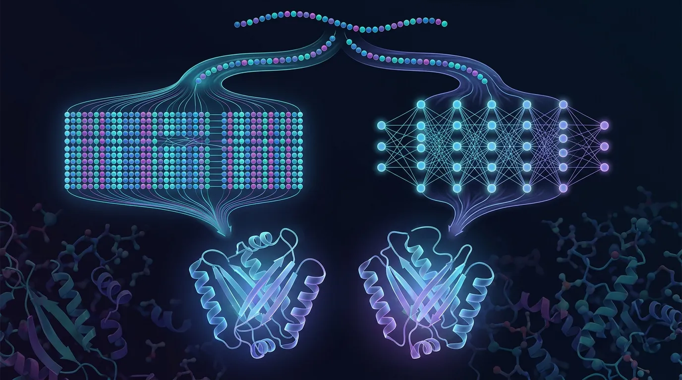
The newest class of methods skips the "find a characterized relative" step and predicts function directly, learning the sequence-to-function mapping from the whole annotated corpus.
Protein language models (the ESM family, and others) train on hundreds of millions of sequences and produce per-residue embeddings that encode a surprising amount of functional signal. Those embeddings feed classifiers that predict GO terms, EC numbers, and localization — without needing a detectable homolog, because the model has internalized regularities across sequence space.
Structure-based predictors go further by reasoning over the fold. DeepFRI combines language-model sequence features with a graph convolutional network over the residue-contact graph to predict GO terms and EC numbers — and, usefully, it highlights the residues most responsible for a predicted function, giving you candidate sites to test rather than a bare label.
Where ML predictors break
- Biased toward what's already annotated. These models learn from the annotated corpus, so they're strongest on well-represented function classes and weakest on the novel and rare — exactly the orphans you care about most.
- The output is a probability, not a fact. A moderate score on a broad term ("catalytic activity," "binding") is rarely actionable. Read the specific, high-confidence terms; discount the vague ones.
- Confidently wrong is possible. With no homolog to anchor it, a model can assign a crisp, plausible, incorrect function to a genuinely novel protein. Confidence is not correctness.
Used well, ML predictors are the best hypothesis generators for proteins with no usable homolog; used naively — copying the top GO term into your annotation — they manufacture false certainty.
Read the Structure You Already Have
Before you order a single reagent, the predicted model itself carries functional clues, if you read it with discipline.
- Pockets and clefts. A deep cavity lined with conserved residues is a candidate binding or active site, and its shape constrains what could bind — but a pocket is an invitation, not a mechanism, and plenty of clefts bind nothing.
- Catalytic geometry. A constellation of residues held in a precise arrangement (a Ser-His-Asp triad, a metal-coordinating His/Glu cluster) is a mechanistic hint no single-residue conservation score would reveal.
- Membrane spans and disorder. A run of hydrophobic helices crossing a slab flags an integral membrane protein; long low-pLDDT stretches are usually intrinsically disordered regions that carry interaction motifs and PTM sites — functional elements, not folding failures.
- Trust the confidence tracks. Interpret pockets and geometry only where pLDDT is high; elsewhere the coordinates are guesses and your "pocket" may be an artifact. The PAE matrix tells you which domains are confidently positioned relative to each other — critical before reasoning about an inter-domain active site.
The Traps That Cost You Months
Most function-prediction disasters aren't exotic. They come from four recurring mistakes.
1. Transferring from a moonlighting or misannotated neighbor. Your top hit is a moonlighting protein and you inherit its secondary function — or it was itself annotated by homology to something annotated by homology to an original guess. Either way you've built on sand. The fix: transfer preferentially from experimentally characterized, manually curated entries (Swiss-Prot reviewed, not TrEMBL automatic), and treat annotation provenance as data.
2. Confusing catalytic residues with binding residues. A conserved residue in a pocket might coordinate a metal, stabilize the fold, or bind substrate without being catalytic — catalytic residues are a specific subset with specific geometry. "Functionally important" is not "performs the reaction," and designing a mutagenesis experiment on the wrong assumption wastes a round of cloning.
3. Annotation error propagation. The systemic trap. In an audit of 37 enzyme families across Swiss-Prot, GenBank NR, TrEMBL, and KEGG, misannotation of molecular function was serious in every database except the manually curated Swiss-Prot — above 80% in the most error-prone families in the automatic resources. Each wrong entry seeds the next automated annotation, so transferring function from "the database" often means copying an error photocopied thousands of times.
4. Betting on a single line of evidence. Homology says one thing, the structural hit another, the ML predictor a third, and you pick whichever confirms your hope. The discipline is the opposite: treat agreement across independent methods as signal, disagreement as a flag to investigate — not to cherry-pick.
How Hard Is This, Really? A Reality Check
Worth stating plainly, because the tooling can create an illusion of solvedness: function prediction is not solved for novel proteins.
The CAFA community experiment evaluated 54 methods on 866 proteins across 11 organisms in a blind, time-separated benchmark. The best methods clearly beat a naive BLAST baseline — but performance stayed well short of reliable, complete annotation, molecular-function prediction was easier than the broader biological-process terms, and the hardest targets (least prior information) were where every method struggled most.
That's the honest frame for everything above: these tools are excellent at generating ranked hypotheses and poor at delivering certainty on a genuine orphan. Plan experiments accordingly.
Case Study: A "Domain of Unknown Function" That Turned Out to Be a Metal-Dependent Hydrolase
Problem. A team pulled a hit from an environmental metagenome — a ~280-residue protein enriched in a sample with unusual degradative activity. AlphaFold returned a clean model, pLDDT 89 in the core, with a promising cleft. BLAST against Swiss-Prot found nothing above 28% identity, and the best hits were themselves uncharacterized. The obvious next step — order a substrate panel and start screening — meant guessing which of a hundred assays to run, at real cost each.
Analysis. They worked the ladder instead of guessing. InterPro placed the protein in a Pfam family flagged as a domain of unknown function — real family, no known activity. Foldseek against the PDB then matched the fold, despite sub-30% sequence identity, to a characterized metallohydrolase superfamily. A structure-based function predictor independently returned "hydrolase activity" and "metal ion binding" among its top GO terms and flagged a cluster of cleft residues — which, read off the model, formed a plausible di-metal coordination geometry: three histidines and an aspartate, arranged the way a metal-dependent hydrolase active site is. Four independent lines — profile family, structural homology, ML prediction, active-site geometry — converged on one hypothesis, not the "unknown" the sequence databases offered.
Solution. Instead of a hundred-substrate fishing expedition, the team ran a targeted test: a small panel of ester and amide substrates for that hydrolase class, with and without a metal chelator to confirm metal dependence, plus an alanine mutant of one predicted metal-coordinating histidine as a mechanistic control.
Outcome. The wild type showed chelator-sensitive hydrolysis on a subset of the panel; the histidine mutant lost most of the activity — consistent with the predicted active site. Months of unfocused screening became one well-scoped experiment, because triage narrowed a hundred assays to a handful and named the control that would prove them wrong. The prediction didn't replace the experiment; it made it cheap and decisive.
A Practical Workflow: From Model to Testable Hypothesis
Run the methods in order of specificity, and stop escalating when you have convergent, high-confidence evidence.
START: You have a sequence (and ideally a predicted structure).

1. HOMOLOGY (BLAST / DIAMOND vs curated databases)
   │
   ├─ Strong hit (>40% id, good coverage) to an EXPERIMENTALLY
   │   CHARACTERIZED, CURATED entry?
   │     └─ YES → Transfer EC / GO / active-site residues.
   │              Verify provenance, then you're largely done.
   │
   └─ Weak / twilight-zone hit, or hit to another uncharacterized
       protein? → escalate.

2. PROFILE METHODS (InterPro / Pfam / HMMER)
   │  Get domain architecture + family + associated GO.
   ├─ Family is a DUF, or a superfamily with many activities?
   │     └─ Architecture known, activity not → escalate.

3. STRUCTURE SEARCH (Foldseek vs PDB/AFDB; DALI to scrutinize)
   │  Use your predicted model as the query.
   ├─ Strong fold match to a characterized protein?
   │     └─ Functional LEAD (remember: fold ≠ function) → carry forward.

4. ML PREDICTORS (protein language model / DeepFRI-style)
   │  Get ranked GO / EC hypotheses + candidate functional residues.
   │  Keep the specific, high-confidence terms; discard the vague ones.

5. READ THE STRUCTURE
   │  Pockets, catalytic geometry, TM spans, disorder — only where
   │  pLDDT/PAE support it.

6. TRIANGULATE
   │  Require ≥2 INDEPENDENT lines to agree before you believe a
   │  function. Disagreement = investigate, don't cherry-pick.

7. DESIGN THE CHEAPEST DISCONFIRMING EXPERIMENT
      A targeted assay + a mechanistic control (e.g., active-site
      mutant) that would prove the hypothesis WRONG if it's wrong.
Pre-experiment checklist:
- Homology hits are to curated, experimentally characterized entries — not automatic annotations or other orphans.
- Structural search run on the predicted model, not just sequence; domain architecture mapped.
- ML predictions filtered to specific, high-confidence terms; pockets and geometry read only in high-pLDDT regions.
- At least two independent methods agree on the proposed function.
- Catalytic vs. binding residues distinguished before designing mutants.
- The planned experiment includes a control that could falsify the hypothesis.
The Economics of Guessing Function Wrong
Getting function wrong is not a small error — it silently redirects an entire program. The cost isn't the compute; it's the wet-lab work built on a bad premise.
| Failure | What it looks like | Real cost |
|---|---|---|
| Wrong EC class | You assay for hydrolase activity; it's a transferase. Every well reads negative. | You conclude "inactive" and shelve a good protein — or burn a 50–100 substrate screen chasing the wrong chemistry. |
| Function from a misannotated homolog | Inherited annotation is a photocopied database error. | Weeks to months of assays for an activity the protein never had. |
| Moonlighting confusion | You test the neighbor's secondary function. | A real function exists, but you validated the wrong one and reported it. |
| Binding vs. catalytic mixup | Alanine-scan the wrong residues. | A full round of cloning, expression, and assay yields uninterpretable data. |
| Wrong target selection | A mis-scoped protein enters a discovery program as the lead. | Months to a year and six figures before the negative result lands. |
The pattern is consistent: minutes of computational triage at the start determine months of experimental work downstream. The expensive mistake is never the prediction — it's committing bench resources to an unverified guess. A day spent triangulating function and designing one disconfirming experiment is the cheapest insurance in the campaign.
Bottom Line
A predicted structure is a scaffold, not an answer. Function is a stack of separate questions — EC number, GO terms, family, localization, topology, binding residues, PTMs — and no single method answers them reliably on a novel protein. Work the ladder in order of specificity: homology first (trusting only curated, characterized hits), profiles for domain architecture, structural search when sequence goes dark, ML predictors for direct hypotheses, and the model itself for pockets and geometry. Then triangulate — demand agreement across independent lines — and design the cheapest experiment that could prove you wrong. The databases are full of confident errors; discipline about provenance and convergence is what keeps you off the months-long detour.
How Orbion Helps
Everything above is a multi-tool scavenger hunt: BLAST in one window, InterPro in another, Foldseek in a third, an ML predictor in a fourth — then the hard work of reconciling them. Orbion's Characterization module runs that convergence in one place, so you read a consolidated functional hypothesis instead of stitching one together by hand.
Paste a sequence and the Overview opens with an AI literature scan and a homology search across 700M+ proteins (UniProt + PDB) run entirely within Orbion infrastructure — your sequence is never sent to a third-party service. Then the Astra models each answer a different piece of the function stack, from sequence and the predicted model:
- AstraROLE predicts the functional core — EC numbers, GO terms, pathways, and protein categories — the "what reaction / what process" layer structure alone can't give you.
- AstraBIND predicts ligand binding sites and active-site residues — turning "there's a pocket here" into candidate positions you can test.
- AstraSUIT predicts classification, host association, membrane type, subcellular location, cofactors, and quaternary structure — the localization and context layer.
- AstraUNFOLD predicts transmembrane helix topology, per-residue disorder, and amyloid propensity — reading membrane spans and disordered functional elements off the sequence.
Around them, AlphaFill transplants ligand and cofactor annotations from homologous PDB structures onto your model, the PAE Insight Engine flags domain boundaries and hinges, and where curated data exists Orbion validates predictions against UniProt — so you see where the model agrees with the record and where it's extrapolating.
The framing is the one this article argues for: triage and hypothesis generation, not a replacement for experiments. Orbion gives you a consensus functional picture and the specific residues and assays worth testing — so the experiment you run is the decisive one, not the tenth guess. Start with your orphan sequence at orbion.life.
References & sources
- Jumper, J. et al. Highly accurate protein structure prediction with AlphaFold. Nature 596:583–589 (2021). doi:10.1038/s41586-021-03819-2 — The structure-prediction breakthrough that created the "structure without function" gap this article addresses.
- Altschul, S. F. et al. Gapped BLAST and PSI-BLAST: a new generation of protein database search programs. Nucleic Acids Research 25:3389–3402 (1997). doi:10.1093/nar/25.17.3389 — The workhorse of homology-based function transfer and the source of its twilight-zone limits.
- Eddy, S. R. Accelerated profile HMM searches. PLoS Computational Biology 7:e1002195 (2011). doi:10.1371/journal.pcbi.1002195 — The profile-HMM search engine (HMMER3) behind Pfam that reaches into the twilight zone pairwise search can't.
- van Kempen, M. et al. Fast and accurate protein structure search with Foldseek. Nature Biotechnology 42:243–246 (2024). doi:10.1038/s41587-023-01773-0 — Structure-based search that lets a predicted model be the query when sequence homology fails.
- Gligorijević, V. et al. Structure-based protein function prediction using graph convolutional networks. Nature Communications 12:3168 (2021). doi:10.1038/s41467-021-23303-9 — DeepFRI: predicts GO/EC from sequence and structure and highlights functionally important residues.
- Ashburner, M. et al. Gene Ontology: tool for the unification of biology. Nature Genetics 25:25–29 (2000). doi:10.1038/75556 — Defines the GO framework (molecular function, biological process, cellular component) that function predictors target.
- Schnoes, A. M. et al. Annotation error in public databases: misannotation of molecular function in enzyme superfamilies. PLoS Computational Biology 5:e1000605 (2009). doi:10.1371/journal.pcbi.1000605 — Quantifies database misannotation and error propagation across four major public databases.
- Jeffery, C. J. Moonlighting proteins. Trends in Biochemical Sciences 24:8–11 (1999). doi:10.1016/S0968-0004(98)01335-8 — The foundational account of single proteins with two unrelated functions — a core homology-transfer trap.
- Radivojac, P. et al. A large-scale evaluation of computational protein function prediction. Nature Methods 10:221–227 (2013). doi:10.1038/nmeth.2340 — The CAFA community benchmark showing how far function prediction still is from solved, especially for orphans.