You mix purified receptor, MSP1D1, and POPC at the ratio that worked in a paper. After overnight detergent removal, the tube is clear. The SEC trace is not: one peak in the void, a broad shoulder across half the column, and a late peak that looks exactly like empty nanodiscs. Nothing visibly precipitated, yet almost none of the target is in a homogeneous protein-loaded disc.
Nanodisc assembly is self-organization with several coupled variables. Protein, scaffold, lipid, and detergent must cross a narrow concentration window while detergent is removed at the right rate. A published ratio is a starting point, not a transferable recipe, because target footprint, oligomeric state, bound detergent, native lipid, scaffold length, and lipid packing all change the geometry.
This guide treats reconstitution as a diagnostic workflow: define the particle you need, screen ratios at small scale, control detergent removal, separate loaded from empty discs, and use the failure pattern to choose the next experiment.
Key Takeaways
- Start with monodisperse, functional protein. Nanodiscs preserve a good input more reliably than they rescue an aggregated detergent sample.
- Optimize three molar ratios together: target:MSP, lipid:MSP, and detergent:adsorbent. The correct values depend on scaffold and target footprint.
- Use a small matrix, not one literature ratio. Three target:MSP ratios crossed with three lipid:MSP ratios usually teaches more than a large single batch.
- Detergent removal is the assembly trigger. Too fast promotes aggregation and liposomes; too slow leaves incomplete or detergent-rich particles.
- SEC alone cannot prove occupancy. Combine chromatogram shape with SDS-PAGE, target-specific affinity enrichment, native mass/SEC-MALS where feasible, and negative-stain EM or another particle readout.
- Diagnose empty discs, oversized discs, aggregation, and lost function separately. They have different causes and opposite fixes.
What Successful Nanodisc Assembly Looks Like
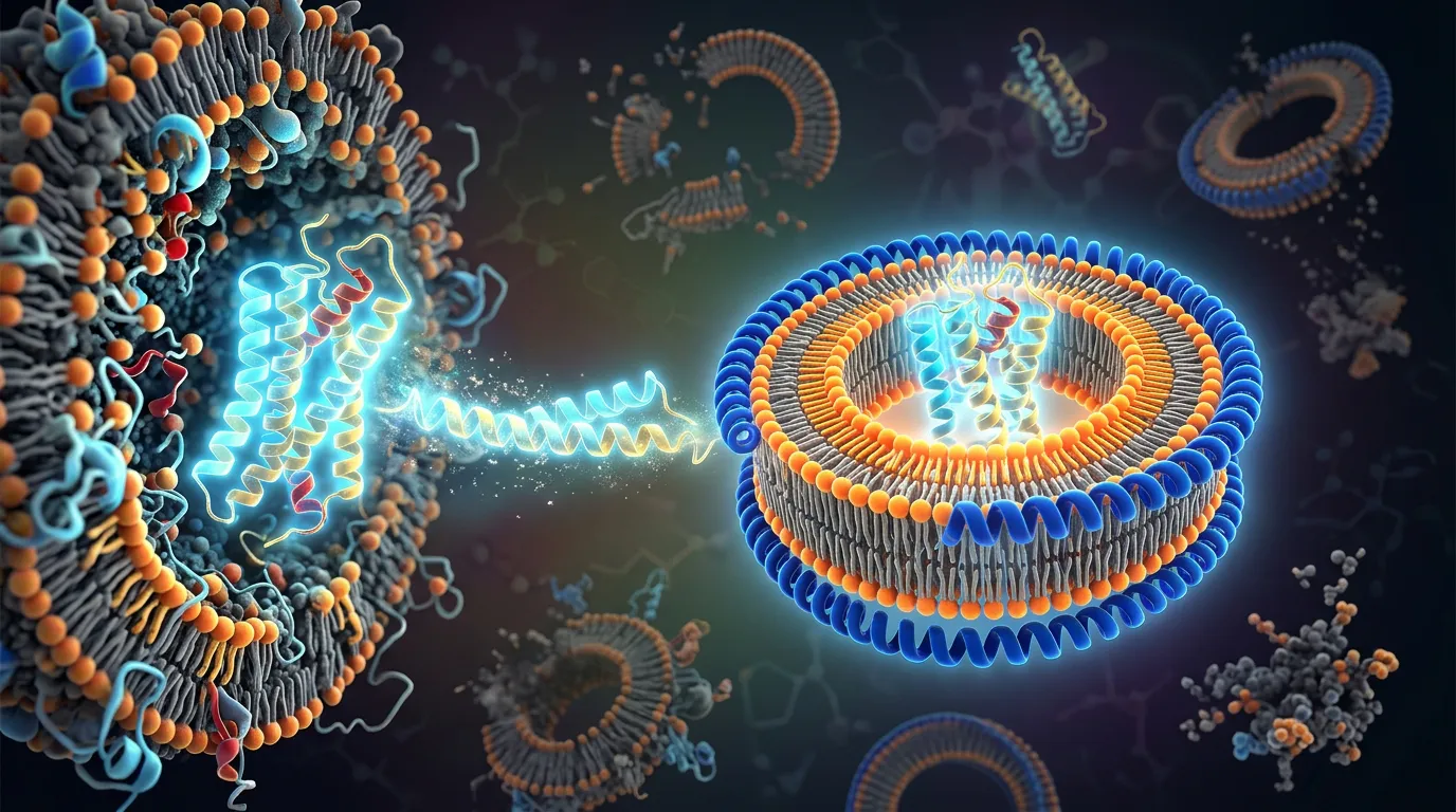
An MSP nanodisc is a patch of phospholipid bilayer wrapped by two amphipathic membrane scaffold protein (MSP) molecules. The target sits inside the bilayer patch; the scaffold shields the lipid edge from water.
A useful protein-loaded preparation should meet four criteria:
- Defined particle size. A dominant, reasonably symmetric SEC peak appears at the expected hydrodynamic size.
- Correct composition. The peak contains both target and MSP at a reproducible ratio.
- Controlled occupancy. The sample is enriched for the desired number of target molecules per disc rather than a mixture of empty, singly loaded, and multiply loaded particles.
- Retained function. Binding, enzymatic, or conformational activity survives reconstitution.
The visual absence of precipitation addresses none of those criteria.
Step 0: Define the Particle Before Choosing the Scaffold
Write down the intended endpoint:
- monomeric receptor for binding assays;
- oligomeric transporter for cryo-EM;
- receptor–G-protein complex;
- lipid-dependent enzyme;
- single-molecule construct with a biotinylated scaffold;
- donor–acceptor geometry for spectroscopy.
Then estimate the membrane footprint of the full assembly, not just the monomer. A scaffold chosen for a compact seven-transmembrane receptor may be too small for a receptor–arrestin complex or a dimeric transporter.
| Scaffold family | Approximate disc regime | Typical use | Common failure if undersized |
|---|---|---|---|
| MSP1D1ΔH5 | Small | small monomeric proteins, NMR-oriented work | target excluded or distorted |
| MSP1D1 | Standard ~10 nm class | many monomeric receptors and transporters | crowding for large complexes |
| MSP1E1 / MSP1E3D1 | Intermediate/large | larger proteins and partner complexes | more empty discs and lipid demand |
| MSP2N2 and larger systems | Large | oligomers and large assemblies | broad size distribution if lipid ratio is not retuned |
Scaffold diameter is not a cosmetic variable. The bilayer patch must accommodate the target plus an annulus of lipids without forcing the MSP belt into an unstable geometry.
For a broader comparison with detergent micelles and polymers, see lipid nanodiscs vs detergent micelles vs SMALPs.
Step 1: Qualify Every Input
Target protein
Use freshly purified or validated frozen material with:
- a dominant non-void SEC peak;
- minimal time-dependent aggregation;
- known concentration by a method compatible with detergent;
- retained ligand/substrate response if measurable;
- intact mass or a convincing full-length band;
- the lowest practical free-detergent load.
If the target is already heterogeneous, isolate the best peak immediately before reconstitution. Do not pool shoulders to improve yield.
MSP
MSP should be highly pure, monodisperse, and free of persistent oligomers. Confirm concentration carefully; a 20% MSP error moves every stoichiometry in the screen. If both MSP and target carry His tags, plan how you will separate empty discs from loaded discs. An untagged scaffold or an orthogonal target tag often simplifies enrichment.
Lipid
Use fresh, well-defined stocks. Oxidized unsaturated lipids can change assembly and function. Dry lipid films thoroughly, resuspend above the main lipid’s phase-transition temperature, and solubilize consistently—often with sodium cholate—until the stock is clear.
For mixtures, prepare a master stock so every ratio condition receives the same composition. Do not pipette tiny volumes from separate lipid stocks into each tube.
Bio-Beads or other adsorbent
Wash detergent-removal resin exactly and remove storage contaminants. Hydrate and equilibrate it in reconstitution buffer. Bio-Beads that carry methanol or inconsistent water content make the detergent-removal rate irreproducible.
Step 2: Choose Lipid Composition With a Hypothesis
POPC is a common neutral starting lipid because it is fluid at room temperature and easy to handle. It is not a universal native mimic.
| Target context | Rational first additives | Why |
|---|---|---|
| Bacterial inner-membrane transporter | POPE/POPG or polar lipid extract | introduces native-like PE and anionic lipid |
| GPCR | POPC with cholesterol or CHS-compatible strategy | sterol can stabilize receptor states |
| Mitochondrial respiratory protein | cardiolipin-containing mixture | cardiolipin may be structural or functional |
| Peripheral/basic membrane-binding region | defined anionic lipid fraction | controls electrostatic recruitment |
| Unknown target | simple PC system plus one native-like mixture | separates assembly chemistry from lipid dependence |
Keep the first matrix interpretable. If you vary scaffold, lipid composition, ratio, detergent-removal rate, ligand, and temperature simultaneously, a good particle teaches you nothing transferable.
Start with one simple composition. After assembly is reproducible, screen functional lipids.
Step 3: Calculate Ratios in Moles
Nanodisc recipes are molar recipes. Convert every input into moles before mixing.
For each component:
moles = mass / molecular weight
For lipid mixtures, use a weighted average molecular mass or calculate each lipid separately. Record the detergent contributed by the target stock and the lipid stock; it controls how much adsorbent you need.
A rational first matrix
Choose three target:MSP ratios and three lipid:MSP ratios around a published starting point for the scaffold. For a monomeric target in MSP1D1, a useful exploratory design is:
- target:MSP = 1:2, 1:5, 1:10;
- lipid:MSP = one lower, one nominal, and one higher condition around the scaffold’s established bare-disc ratio.
That produces nine small reactions. The exact lipid numbers depend on lipid identity and scaffold; use the MSP protocol rather than copying a ratio across scaffold variants.
Why include excess MSP? A target:MSP ratio such as 1:10 can improve the probability that each target enters a separate disc, at the cost of many empty particles. That can be acceptable if the target has an orthogonal affinity handle for post-assembly enrichment.
Why include a lower-MSP condition? If excess empty discs are the dominant problem and the target tolerates closer packing, lower MSP can improve loaded-particle yield. It also raises the risk of multi-occupancy and aggregation.
Step 4: Mix in the Correct Order
A robust default order is:
- Combine detergent-solubilized target with solubilized lipids.
- Incubate gently so the protein exchanges into the chosen lipid environment.
- Add MSP.
- Incubate again before initiating detergent removal.
The target-first lipid incubation is not universal, but it reduces abrupt exposure of a fragile protein to a rapidly assembling scaffold/lipid mixture. Keep all reactions at a temperature compatible with both protein stability and lipid fluidity. If the main lipid is below its phase-transition temperature, assembly can become slow or heterogeneous.
Avoid vortexing after the target is added. Use gentle inversion or slow rotation.
Step 5: Remove Detergent Deliberately
Detergent removal drives self-assembly. Bio-Beads adsorb free detergent; as the concentration falls, lipids form bilayer patches and MSP closes around the edges.
A published practical workflow uses approximately 20 mg Bio-Beads per 100 µL reaction for an initial two-hour incubation, followed by fresh beads and gentle mixing overnight. That is a starting schedule, not a universal capacity rule, because adsorption differs among DDM, LMNG, cholate, GDN, and mixtures (Miehling et al., 2022).
If removal is too fast
- target aggregates before a disc closes;
- lipids form vesicles or large sheets;
- MSP precipitates with lipid;
- the reaction develops a large void-volume peak.
If removal is too slow or incomplete
- particles remain detergent-rich;
- SEC peaks broaden;
- scaffold/lipid assemblies do not close uniformly;
- downstream assays retain detergent artifacts;
- loaded discs may dissociate during dilution.
Control the rate
- add adsorbent in two or three stages;
- use dialysis plus adsorbent for gentler removal;
- lower temperature for fragile proteins, while keeping lipids fluid;
- reduce starting detergent load before assembly;
- choose a more removable detergent for the final purification step.
LMNG and GDN can stabilize a target beautifully and resist removal. If reconstitution repeatedly fails from an LMNG stock, exchange the target into DDM or another validated, more removable detergent before assembly rather than adding more Bio-Beads blindly.
Step 6: Clarify Without Throwing Away the Answer
After detergent removal:
- separate the beads gently;
- clarify the sample with a short spin to remove visible particles;
- keep aliquots of total reaction, pellet, and supernatant;
- filter only if needed and record recovery.
If you analyze only the final supernatant, you cannot tell whether a low-yield SEC peak reflects poor assembly, target precipitation, adsorption to the beads, or loss on the filter.
Run SDS-PAGE on the mass-balance fractions. A target that disappeared onto Bio-Beads needs a different removal schedule; a target in the pellet needs a different ratio or input condition.
Step 7: Separate Loaded and Empty Nanodiscs
SEC separates aggregates, large assemblies, correctly sized discs, and small free components. It often does not fully separate empty from target-loaded discs when their hydrodynamic sizes overlap.
Use an occupancy strategy:
- His-tagged target plus untagged MSP, followed by IMAC enrichment;
- biotinylated or FLAG-tagged target with compatible affinity capture;
- target-specific ligand resin when function is preserved;
- density or ion-exchange differences for specialized systems.
Then run a final polishing SEC.
For a step-by-step ABC-transporter reconstitution, Bao and Duong mixed detergent-solubilized MalFGK2 with MSP and lipid, removed detergent with adsorbent, and separated particles by centrifugation and SEC; their protocol emphasizes that assembly and purification are distinct stages (Bao & Duong, 2012).
Step 8: Prove Particle Identity and Function
Use at least three orthogonal readouts.
Composition
SDS-PAGE or capillary electrophoresis should show target and MSP in the expected fractions. Densitometry is approximate because staining differs between proteins; use it to compare conditions, not to claim exact stoichiometry.
Size and homogeneity
- analytical SEC;
- negative-stain EM;
- DLS as a quick polydispersity check;
- SEC-MALS where detergent/lipid modeling is appropriate;
- mass photometry or native MS for compatible systems.
Function
- ligand binding;
- ATPase or enzymatic activity;
- conformational antibody binding;
- transport/flux only after transfer to a compartment-forming system if required.
Nanodiscs provide a bilayer patch but not an enclosed lumen. They cannot directly replace proteoliposomes for transport gradients or channel flux.
Troubleshooting by SEC Signature
Failure A: Large void-volume peak
Likely causes
- target was aggregated before assembly;
- disc is too small for the target;
- too little MSP;
- too little lipid;
- detergent removal was too fast;
- target:target occupancy is too high.
Next experiment
Use a larger scaffold, increase MSP, increase lipid modestly, reduce target loading, stage Bio-Bead addition, and requalify the input target by SEC.
Failure B: Broad peak spanning loaded and empty-disc regions
Likely causes
- lipid:MSP ratio is off;
- mixed occupancy states;
- incomplete detergent removal;
- broad target oligomer distribution.
Next experiment
Narrow the ratio matrix around the best condition, add affinity enrichment for loaded discs, and measure target oligomer state before reconstitution.
Failure C: Dominant empty-disc peak, little target
Likely causes
- MSP is in large excess;
- target precipitated or adsorbed to beads;
- target did not exchange out of its detergent micelle;
- disc is too small or lipid composition is unfavorable.
Next experiment
Lower MSP relative to target, track the target mass balance, exchange the detergent, and test a larger scaffold. If the target has an orthogonal tag, accept excess empty discs and enrich loaded particles rather than sacrificing monodispersity.
Failure D: Correct-looking disc peak, no activity
Likely causes
- lipid composition is wrong;
- target orientation/occupancy is heterogeneous;
- required ligand, cofactor, or partner was lost;
- detergent exposure inactivated the target before assembly;
- the assay requires a sealed compartment.
Next experiment
Compare activity before and after reconstitution, screen one lipid variable at a time, include ligand/cofactor during assembly, and move to proteoliposomes if a gradient is required.
Failure E: Sample works immediately, aggregates after concentration or freezing
Likely causes
- discs collide or stack at high concentration;
- free lipid/detergent changes during concentration;
- freeze-induced phase behavior damages the bilayer;
- buffer or cryoprotectant is incompatible.
Next experiment
Concentrate more slowly, use a higher-MWCO device validated for discs, compare fresh versus frozen material, and optimize salt/cryoprotectant with a functional readout.
A Nine-Reaction Rescue Matrix
When the first attempt fails, do not change everything. Run:
| Low lipid:MSP | Nominal lipid:MSP | High lipid:MSP | |
|---|---|---|---|
| Target:MSP 1:2 | A1 | A2 | A3 |
| Target:MSP 1:5 | B1 | B2 | B3 |
| Target:MSP 1:10 | C1 | C2 | C3 |
Keep scaffold, lipid composition, temperature, ligand, buffer, and removal schedule constant. Rank conditions by:
- target recovery after bead removal;
- void-volume fraction;
- symmetry of the disc peak;
- target:MSP signal in that peak;
- retained activity.
Then run a second matrix around the best ratio, changing either scaffold size or lipid composition—not both.
Worked Example: Rhodopsin Shows Why Ratios Are Target-Specific
Rhodopsin has been assembled into MSP nanodiscs with several different recipes depending on scaffold, lipid, and experimental aim. In one protocol, rhodopsin, MSP, and POPC/POPG were mixed at a 0.1:1:75 molar ratio, detergent was removed overnight with Bio-Beads at 4 °C, and particles were polished by SEC. The resulting discs supported membrane-like photointermediates (Tsukamoto et al., 2011).
Another MSP1E3D1 workflow used an excess of scaffold and lipid to favor monomeric receptor occupancy and separated a small aggregate fraction by SEC (Bayburt & Sligar, 2010).
These are not conflicting recipes. They illustrate why the target state, scaffold, lipid, and occupancy objective define the ratio.
The Economics of Reconstitution
| Choice | Material cost | Time cost | Information value |
|---|---|---|---|
| One large literature-ratio batch | High | Low initially | Poor if it fails |
| Nine-condition microscreen | Moderate | 2–3 days | High; maps the ratio landscape |
| Immediate broad lipid screen | High | High | Low until assembly itself is controlled |
| Scaffold-size comparison after ratio screen | Moderate | 2–3 days | High when crowding is suspected |
The scarce resource is purified membrane protein. Use it to measure a small design space, not to maximize the volume of an unvalidated condition.
Bottom Line
Nanodisc reconstitution succeeds when a qualified target, an appropriately sized scaffold, a compatible lipid mixture, and a controlled detergent-removal schedule converge on the same particle. Start with a molar matrix, preserve a complete mass balance, enrich loaded discs, and require function—not just a clear tube or an SEC peak.
The failure pattern is a map. Empty discs call for a different occupancy strategy; void-volume material calls for gentler assembly or more space; a homogeneous inactive disc calls for biological context, not prettier chromatography.
Planning Nanodisc Reconstitution With Orbion
Orbion’s Characterization module maps membrane topology, lipid-facing helices, disorder, binding sites, cofactors, and confidence onto the target. Design helps choose boundaries, topology-compatible tags, and constructs sized for the intended assembly. Bench can generate a reconstitution screen with explicit target:MSP:lipid ratios, detergent-removal variables, and QC acceptance criteria.
That turns a copied recipe into a target-specific experiment.
References
- Denisov, I. G. et al. Directed self-assembly of monodisperse phospholipid bilayer Nanodiscs with controlled size. Journal of the American Chemical Society 126, 3477–3487 (2004). doi:10.1021/ja0393574
- Bao, H. & Duong, F. A step-by-step method for the reconstitution of an ABC transporter into nanodisc lipid particles. Journal of Visualized Experiments 66, e3910 (2012). Full text
- Miehling, J. et al. Reconstitution of membrane proteins into platforms suitable for biophysical and structural analyses. Methods in Molecular Biology 2507, 183–205 (2022). Full text
- Bayburt, T. H. & Sligar, S. G. Membrane protein assembly into Nanodiscs. FEBS Letters 584, 1721–1727 (2010). doi:10.1016/j.febslet.2009.10.024
- Tsukamoto, H. et al. Rhodopsin in nanodiscs has native membrane-like photointermediates. Biochemistry 50, 5086–5091 (2011). Full text
- Ritchie, T. K. et al. Reconstitution of membrane proteins in phospholipid bilayer nanodiscs. Methods in Enzymology 464, 211–231 (2009). doi:10.1016/S0076-6879(09)64011-8