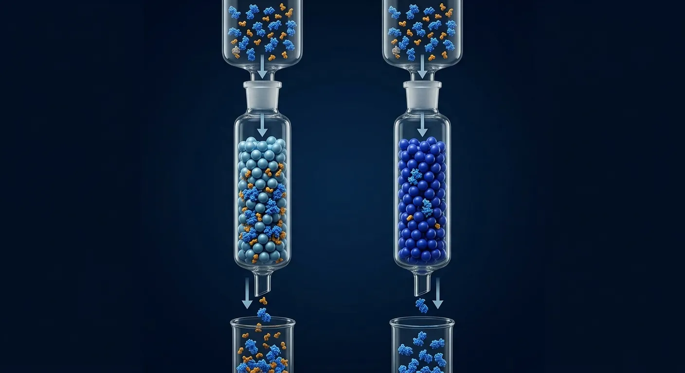
The fusion expressed, the affinity step worked, and then tag removal turned one clean band into three. Protease choice is often made from a vector map before anyone considers the target’s N terminus, solvent accessibility, temperature tolerance, internal sequence liabilities, or the downstream method used to remove the protease.
TEV, HRV 3C, thrombin, and SUMO protease solve different problems. The best enzyme is not the one with the most familiar recognition site. It is the one that produces the required product sequence, cleaves under target-compatible conditions, avoids internal cuts, and can be removed without sacrificing the protein.
Key Takeaways
- Start with the desired product termini. SUMO protease can release a native N terminus; TEV and HRV 3C commonly leave one or more engineered residues.
- Specificity is sequence- and structure-dependent. Scan the full target for accessible internal motifs; even highly specific proteases can cleave noncanonical sites.
- Accessibility often limits cleavage. A perfect site buried against the target or resin will react slowly.
- Temperature and buffer compatibility matter. HRV 3C and engineered TEV variants are useful at cool temperatures; thrombin can be fast but less sequence-selective.
- Design protease removal before cleavage. Tagged protease plus subtractive IMAC is convenient only if the released product does not rebind.
- Pilot concentration, enzyme ratio, and time together. Overnight digestion is not an optimization strategy.
The Four Strategies at a Glance
| Protease | Typical recognition logic | Main advantage | Main risk |
|---|---|---|---|
| TEV | extended linear motif, commonly ENLYFQ↓G/S | high specificity, broad use | slow at inaccessible sites; noncanonical internal cuts occur |
| HRV 3C | commonly LEVLFQ↓GP | active at cool temperature, specific | product often retains GP; site context matters |
| thrombin | basic motif around LVPR↓GS | rapid, familiar | broader specificity and internal cleavage risk |
| SUMO protease | recognizes folded SUMO domain and junction | native N terminus possible | requires correctly folded SUMO and a compatible junction |
Recognition sequences shown are common designs, not complete specificity models. Residues flanking the motif and three-dimensional access affect cleavage.
Decision 1: What Must the Final N Terminus Be?
For many biochemical assays, an extra glycine or serine is harmless. For N-terminal processing, degradation signals, cyclization, structural studies, and therapeutic peptides, one residue can change the biology.
If an exact native N terminus is required
SUMO protease is often the strongest option. It recognizes the tertiary structure of SUMO and cleaves at the SUMO–target junction, allowing the first target residue to be native. The Ulp1-based system was developed specifically to pair solubility enhancement with precise cleavage (Malakhov et al., 2004).
The cost is construct dependence: SUMO must fold, and the target junction must be accessible. If the fusion accumulates in inclusion bodies, cleavage requires refolding the SUMO domain first.
If a short scar is acceptable
TEV and HRV 3C offer simple linear sites. TEV commonly prefers glycine or serine immediately after the scissile bond, although P1′ tolerance can be broader than the canonical site suggests (Kapust et al., 2002). HRV 3C designs frequently leave GP at the product N terminus.
If the product is not terminus-sensitive
Thrombin may be convenient, especially when the vector already contains its site. But convenience is not a reason to accept internal cleavage. Scan first.
Decision 2: How Specific Must the Reaction Be?
TEV and HRV 3C are popular because their extended motifs are uncommon. That lowers risk; it does not eliminate it.
Recent work demonstrated unexpected TEV cleavage at noncanonical sequences and showed that local context can create experimentally relevant off-target sites (Hu et al., 2024). Our guide to cryptic protease cleavage explains how to distinguish a protease liability from host-cell degradation.
Thrombin recognizes a shorter, more permissive basic motif and can cut at secondary sites, particularly during long or high-enzyme digests. It is best reserved for targets whose sequence and pilot digest show a clean window between complete junction cleavage and internal damage.
SUMO protease gains specificity by recognizing a folded domain, which makes internal target cleavage less likely. However, a poorly folded or aggregated fusion can be resistant even when the junction sequence is correct.
Decision 3: Is the Site Physically Accessible?
Proteases act on conformations, not vector diagrams. A cleavage site can be hidden by:
- a target terminus packed against the protein core;
- a rigid or too-short linker;
- fusion dimerization;
- adsorption to affinity resin during on-column cleavage;
- detergent micelles or nanodiscs;
- target aggregation at the cleavage concentration.
Diagnostic experiments:
- compare cleavage in native and mildly denaturing conditions;
- add 3–8 flexible residues between the protease site and target;
- test cleavage in solution rather than on-column;
- lower protein concentration;
- move the fusion to the opposite terminus;
- confirm that the uncleaved band is intact fusion, not a modified species.
Our linker selection guide covers the spacing problem in more detail.
Decision 4: What Conditions Can the Target Survive?
Temperature
Standard TEV can lose efficiency at low temperature, though engineered variants improve solubility and activity. HRV 3C is widely used for cold cleavage and can suit aggregation-prone targets. Thrombin often works quickly at room temperature, which may accelerate target damage. SUMO protease performance depends on the enzyme preparation and fusion state.
Reducing environment
TEV is a cysteine protease and usually benefits from a reducing agent. HRV 3C also uses a catalytic cysteine. If the target contains required disulfides, find the lowest reductant exposure that preserves protease activity without scrambling the product.
Salt and detergent
Moderate salt is often tolerated, but high salt, ionic detergents, or chaotropes can suppress cleavage or unfold the recognition context. Membrane-protein fusions require a detergent compatibility pilot rather than a generic overnight recipe.
pH
Each enzyme has a useful pH window. Choose a shared window with the target and verify pH after concentrated protein, glycerol, or high salt is added.
Build the Purification Around Protease Removal
A common architecture is:
His6–fusion–site–target + His-tagged protease → released target + tagged fusion + tagged protease
Subtractive IMAC then retains the fusion and protease while the untagged target flows through.
This elegant scheme fails when:
- the target has a native metal-binding patch and rebinds;
- the target retains a His-rich scar;
- imidazole or metal damages the target;
- cleavage is incomplete and uncleaved fusion contaminates the flow-through;
- the protease tag is inaccessible.
Alternative removal methods include ion exchange, size exclusion, a protease-specific inhibitor/resin, or a second orthogonal tag. Model the mass balance before scaling.
A Quantitative Cleavage Screen
Do not optimize one variable at a time with overnight endpoints. Use a small matrix.
Variables
- two protein concentrations;
- three enzyme:substrate mass ratios;
- two temperatures;
- four time points;
- with and without one target-compatible additive if needed.
Readouts
- percentage uncleaved fusion;
- intact target percentage;
- appearance of lower-mass fragments;
- soluble recovery;
- SEC behavior;
- function after protease removal.
Plot intact product versus time. A useful condition reaches near-complete junction cleavage before internal fragments appear. If both curves rise together, changing enzyme amount will not create a clean window; redesign the site or choose a different protease.
Enzyme-by-Enzyme Guidance
TEV
Choose TEV for general-purpose, high-specificity cleavage when a short scar is acceptable and a reducing environment is compatible. Use a soluble engineered variant and include enough linker for access. Check the target for canonical and plausible noncanonical internal sites.
TEV is often the best first choice, but it is not automatically gentle. Long incubations compensate for poor access while increasing oxidation, aggregation, and off-target cleavage.
HRV 3C
Choose HRV 3C when cool-temperature cleavage is important and the GP-like remnant is acceptable. It is common in structural-biology vectors and can be highly efficient in cold rooms. Experimental specificity profiling and substrate studies show that residues around the core motif affect cleavage; verify the exact junction rather than treating the site as a binary string.
Thrombin
Choose thrombin when speed and an established construct matter, the target lacks vulnerable internal motifs, and a pilot digest shows a clean endpoint. Stop the reaction promptly. Avoid adding excess enzyme to force an inaccessible junction.
SUMO protease
Choose SUMO protease when solubility enhancement and a native N terminus are both required. Confirm SUMO folding and plan how to remove the SUMO tag and protease. For insoluble fusions, refolding may become the dominant development problem.
On-Column or In-Solution Cleavage?
On-column cleavage
The tagged fusion stays immobilized while protease releases the target. This can combine cleavage and capture, reduce handling, and keep the target dilute. It works best when the site projects away from the resin and the released target has no affinity for the matrix.
Failure modes include steric occlusion at the bead surface, incomplete diffusion of protease into pores, target rebinding, and long residence under nonideal capture conditions. Because uncleaved fusion remains on the column, apparent purity can look excellent even when yield is poor.
In-solution cleavage
The fusion is eluted, adjusted into a cleavage-compatible buffer, and digested before subtractive purification. This gives better control of enzyme ratio and time and usually improves access. It can also expose a large concentration-dependent solubility drop as the tag is removed.
For a new target, test both at microscale. Choose on-column for a clean, accessible junction and robust target. Choose solution cleavage when kinetics, additives, or product recovery need tight control.
Cleavage Scars Can Change More Than Mass
One or two extra residues may alter N-terminal acetylation, cyclization, degradation, membrane targeting, or crystal contacts. At the C terminus, a remnant can disrupt a peroxisomal targeting sequence, prenylation motif, PDZ-binding motif, or short regulatory tail.
Before accepting a scar:
- map known motifs within ten residues of the terminus;
- check whether the assay uses terminal binding;
- compare native and scarred constructs when structural work is planned;
- confirm the final sequence by intact mass and peptide mapping;
- state the extra residues in methods and reagent records.
For a recombinant enzyme whose active site is distant from the terminus, a glycine may be negligible. For a peptide ligand or signaling domain, it may be the central experimental variable.
How to Stop the Reaction
Cooling or freezing does not always stop proteolysis instantly. A tagged protease can be removed by affinity capture, thrombin can be inhibited or captured with specific reagents, and buffer exchange can remove required cofactors or reductant. Plan a rapid stop compatible with the target.
Take the endpoint sample before and after the stop step. If fragments appear only during overnight storage, the reaction was not truly stopped. For sensitive proteins, run cleavage during the day, sample every hour, and remove the enzyme as soon as the clean window is reached.
Batch Variability and Protease Quality
Protease preparations differ in active fraction, autoproteolysis, tag integrity, and storage history. Define enzyme ratio by active units when possible rather than total mass. Include a control substrate and avoid repeated freeze–thaw cycles.
If the same recipe changes across lots, titrate the new lot against retained reference fusion. A cleavage process is a biochemical reaction and deserves the same incoming-reagent qualification as a chromatography resin.
Troubleshooting Decision Tree
No cleavage
Confirm enzyme activity on a control substrate. Then change access: add linker, lower target concentration, change fusion orientation, or cleave in solution. Verify buffer compatibility and reducing state.
Partial cleavage plateaus
The sample may contain multiple conformations or a modified junction. Compare intact masses, SEC fractions, and native versus mildly denaturing cleavage.
Junction cleavage plus smaller fragments
Map fragments by mass spectrometry or terminal sequencing. Reduce time and enzyme, lower temperature, or switch protease. Do not assume all small bands are host proteolysis.
Target precipitates after cleavage
The fusion was providing solubility or preventing an exposed interface. Cleave at lower concentration, include ligand/cofactor, change salt/pH, or redesign the construct. Our guide to solubility tags addresses this transition.
Protease cannot be removed
Use a differently tagged enzyme, change subtractive resin, or separate by ion exchange/SEC. If the target itself binds IMAC, a His-tagged protease is a poor choice for that workflow.
Activity disappears although the band is intact
Check whether the tag was stabilizing an oligomer, whether the new terminus is correct, and whether reducing agent, metal, imidazole, or the cleavage pH altered the target.
Worked Example: Why More TEV Made the Sample Worse
A 70 kDa fusion reaches 60% cleavage overnight at 4 °C. Increasing TEV fivefold raises cleavage to 90% but creates two target fragments.
A time course shows that junction cleavage is fast for one conformer and slow for another; internal cleavage accumulates after six hours. Adding a six-residue Gly/Ser linker makes the junction accessible, allowing 95% cleavage in two hours with less enzyme and no detectable fragments.
The problem was geometry, not protease potency.
The Economics of Cleavage Design
| Early decision | Cheap test | Expensive error avoided |
|---|---|---|
| exact product terminus | sequence review | remaking material for a terminus-sensitive assay |
| internal-site risk | in-silico scan + pilot digest | losing a scale batch to cryptic cleavage |
| site access | linker/orientation microscreen | escalating protease indefinitely |
| removal strategy | 100 µL subtractive capture | discovering the target rebinds after cleavage |
| post-cleavage solubility | concentration matrix | precipitating the entire purified pool |
Bottom Line
Choose the protease from the final product backward. Define the required terminus, scan for internal liabilities, make the site accessible, match the reaction to the target’s stable buffer, and design removal as part of the same workflow. A short quantitative time course is more informative than an overnight digest at one enzyme ratio.
Designing Tag Removal in Orbion
Orbion Characterize maps termini, disorder, structure confidence, and sequence motifs that affect site accessibility and off-target risk. Design compares fusion orientation, linker length, and cleavage scars. Bench turns the selected strategy into a literature-grounded reaction and purification plan with time-course, recovery, and identity checkpoints.
The right cleavage strategy is the one that delivers the intended protein—not merely a missing fusion band.
References
- Kapust, R. B. et al. The P1′ specificity of tobacco etch virus protease. Biochemical and Biophysical Research Communications 294, 949–955 (2002). doi:10.1016/S0006-291X(02)00574-0
- Hu, J. et al. Unexpected cleavage by tobacco etch virus protease at noncanonical sites. Protein Expression and Purification 217, 106488 (2024). doi:10.1016/j.pep.2024.106488
- Raran-Kurussi, S. et al. Directed evolution of tobacco etch virus protease to enhance its performance in Escherichia coli. Protein Engineering, Design & Selection 30, 791–798 (2017). doi:10.1093/protein/gzx063
- Ullah, R. et al. Activity of the human rhinovirus 3C protease studied in various buffers, additives and detergent solutions for recombinant protein production. PLOS ONE 11, e0153436 (2016). doi:10.1371/journal.pone.0153436
- Malakhov, M. P. et al. SUMO fusions and SUMO-specific protease for efficient expression and purification of proteins. Journal of Structural and Functional Genomics 5, 75–86 (2004). doi:10.1023/B:JSFG.0000029237.70316.52