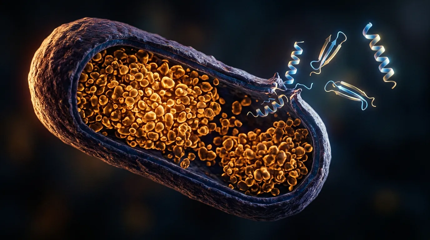
Your SDS-PAGE says the protein expressed beautifully — a fat band at exactly the right molecular weight. Your supernatant says otherwise: it's clear, and your target is sitting in the pellet. It's locked inside inclusion bodies — dense cytoplasmic aggregates of your own overexpressed protein. Weeks of cloning, transformation, and induction to reach one of the most common failure modes in recombinant expression.
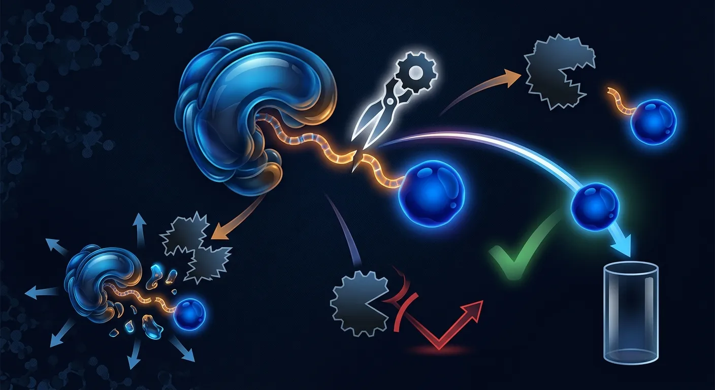
Now you have a decision to make, and it's not "how do I dissolve this pellet?" It's a fork with three roads that have wildly different cost, time, and success profiles: refold the protein out of the inclusion bodies, redesign the construct so it expresses solubly, or re-host — change the strain, compartment, or organism. Pick the wrong road and you can lose a month proving what the sequence would have told you in an afternoon. This guide is about picking the right one.
This is the reactive counterpart to our companion piece on predicting solubility before you express. That one helps you avoid the pellet. This one is for when you're already staring at it.
Key Takeaways
- Inclusion bodies are a fork, not a verdict. Three roads out — refold, redesign, re-host — have very different economics. Choose by what the sequence says, not by whatever's fastest to try first.
- Refolding is the lowest-throughput, most protein-dependent road. Solubilize in 6–8 M urea or 6 M guanidine, then refold by dilution, dialysis, or on-column; recoveries commonly land in the 15–40% range and can be far lower, with no universal protocol.
- If your protein is disulfide-rich or natively secreted, stop fighting the cytoplasm. The E. coli cytoplasm is a reducing environment that can't form stable disulfide bonds — re-host to the periplasm, a disulfide-competent strain (SHuffle/Origami-type), or a eukaryotic host instead of forcing a refold.
- If the trouble is a disordered or aggregation-prone terminus, redesign is the cheapest fix. Truncating a flagged N/C tail or adding a solubility fusion (MBP, SUMO, NusA, TrxA) is one cloning round — not a multi-week refolding campaign.
- Sequence-level predictions pick the road before you burn weeks. Per-residue disorder, amyloid/aggregation propensity, and transmembrane topology tell you whether to truncate, re-host, or refold — and whether refolding even has a chance.
- Sometimes the pellet is the point. Inclusion bodies are near-pure, protease-protected, and one of the few ways to express toxic proteins. Before you "fix" them, ask whether they're already an asset.
First, Confirm It Really Is Inclusion Bodies
Before committing to any road, make sure you're solving the right problem — "in the pellet" is not the same as "in inclusion bodies." A quick fractionation triage on a small culture:
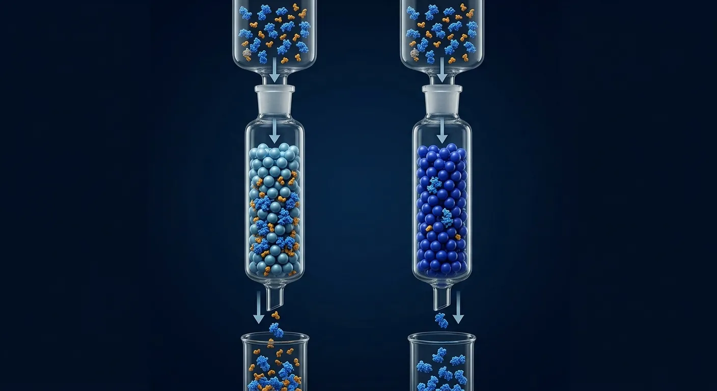
- Lyse and spin. Run supernatant and pellet side by side. A band overwhelmingly in the pellet means insoluble protein — but not yet a diagnosis.
- Rule out membrane association. A protein with transmembrane helices pellets with the membrane fraction and looks "insoluble," but it isn't aggregated — it's in the wrong solvent. Topology prediction flagging transmembrane segments means a detergent/nanodisc workflow, not an inclusion-body one.
- Confirm classic inclusion bodies. They dissolve in strong chaotrope (6–8 M urea, 6 M guanidine hydrochloride) but not mild detergent. If mild detergent releases the protein, suspect membrane partitioning or a loose aggregate.
The rest of this guide assumes confirmed, chaotrope-soluble inclusion bodies of a protein that's supposed to be soluble.
Why Your Protein Went Into the Pellet
Inclusion bodies form when nascent chains aggregate faster than they fold. High-level expression of heterologous proteins in E. coli frequently drives the product straight into them, because over-expression floods the cell with unfolded chains far faster than its chaperone capacity can handle (Baneyx & Mujacic, 2004). Six mechanisms do most of the damage, usually in combination:
- Translation outpaces folding. A strong promoter and high inducer push synthesis faster than folding kinetics allow. Partially folded intermediates, hydrophobic surface still exposed, collide and stick before reaching the native state. This is why "just optimize the IPTG" so often fails — if folding is the bottleneck, more expression makes aggregation worse.
- Exposed hydrophobic and aggregation-prone segments. Short stretches with high intrinsic β-aggregation propensity nucleate amyloid-like assembly between molecules. One or two hotspots buried in the core are harmless; the same segments exposed on a slow-folding intermediate are seeds.
- Intrinsic disorder. Long disordered regions — especially hydrophobic or amyloidogenic ones at the termini — are conformationally promiscuous and frequently drive aggregation in a heterologous host.
- Disulfide bonds that can't form. The E. coli cytoplasm is kept reducing by the thioredoxin and glutaredoxin systems, so a protein that needs disulfides to fold cannot form stable ones there — it misfolds and aggregates. This alone accounts for a large share of secreted proteins that fail in bacterial cytoplasm.
- Missing chaperones, cofactors, or partners. A protein that folds around a metal, a heme, a ligand, or an obligate partner subunit has nothing to fold around when expressed alone; the apo-form is often aggregation-prone.
- Sheer overexpression. Even a well-behaved protein can be forced into inclusion bodies by brute-force expression that overwhelms the folding machinery.
The practical point: the mechanism that put your protein in the pellet is what decides the road out. A disulfide problem and a disordered-terminus problem look identical on a gel and require opposite fixes.
When Inclusion Bodies Are a Feature, Not a Bug
Before spending anything to eliminate them, consider whether inclusion bodies are working in your favor. Sometimes the pellet is the plan:
- Purity. Often 50–90% target protein by mass before a single column — a few washes give enriched starting material a soluble prep can't match.
- Protease protection. Aggregated protein is largely shielded from cytoplasmic proteases, preserving a construct that would otherwise be chewed up.
- Toxic proteins. If your protein kills the host when active and soluble — a nuclease, a protease, a membrane-permeabilizing peptide — sequestering it as an inert aggregate is a feature: high yield of a product that couldn't accumulate any other way, refolded in vitro where toxicity is moot.
- High yield. Inclusion bodies routinely reach gram-per-liter levels, far above what many proteins reach solubly.
If any of these apply, the refold road below isn't a fallback — it's the intended route. Otherwise, read on: you want soluble, folded protein, and the inclusion bodies are in your way.
Three Roads Out: Refold, Redesign, Re-Host
Everything downstream reduces to three strategies. They are not equally good for every protein, and the whole skill is matching the road to the cause.
| Road | What you change | Best when | Throughput | Typical cost |
|---|---|---|---|---|
| Refold | Nothing in the construct — you process the aggregate in vitro | The protein is intrinsically foldable, few/no disulfides, and you want to keep the exact sequence and host | Low (one protein, many condition screens) | Reagents + weeks of optimization |
| Redesign | The construct: truncations, boundaries, solubility fusions, tag placement, induction | The aggregation is driven by a disordered/aggregation-prone region or the protein just needs a folding chaperone-fusion | Medium (parallel constructs, one cloning round) | Cloning + one expression round |
| Re-host | The strain, compartment, or organism | The protein needs disulfides, glycosylation, a eukaryotic chaperone system, or a cofactor the host can't supply | Medium | New vector/strain + one expression round |
The rest of the guide walks each road, then shows how sequence-level predictions tell you which one to take before you commit.
Road 1 — Refold From Inclusion Bodies
Refolding keeps your construct and host untouched and recovers native protein from the aggregate in vitro. It's the right road when the protein is intrinsically foldable and you either need this exact sequence or deliberately expressed into inclusion bodies (toxicity, yield). It's also the lowest-throughput, least predictable road — no universal protocol, and success is strongly protein-dependent (Vallejo & Rinas, 2004). Three stages:
1. Isolate and solubilize
Wash the inclusion bodies (low-urea or detergent washes strip off contaminating membranes and host proteins — where inclusion-body purity pays off), then dissolve the aggregate in strong chaotrope: 6–8 M urea or 6 M guanidine hydrochloride, usually with a reducing agent to break scrambled disulfides. Guanidine is the stronger denaturant for stubborn aggregates; urea is cheaper and more downstream-compatible but can carbamylate proteins if old or heated (Singh & Panda, 2005).
2. Refold by removing denaturant
Lower the denaturant slowly enough that the chain folds instead of re-aggregating. The methods, simplest to most controlled:
- Dilution (drop-wise or pulse into a large volume of refolding buffer): simplest and most scalable, but dilute product needs concentrating afterward.
- Dialysis (gradual removal across a membrane): gentle, good at lab scale, slow.
- On-column / matrix-assisted refolding: bind the denatured, tagged protein to a resin (IMAC for a His-tag), then run a decreasing-denaturant gradient while it's immobilized. Immobilization separates molecules and suppresses the collisions that cause re-aggregation — often the highest-recovery option for aggregation-prone targets.
3. Handle disulfides and additives
Disulfide-bonded proteins need a redox shuffling system — a mix of reduced and oxidized thiols (typically GSH/GSSG at a tuned ratio) so incorrect disulfides can break and re-form until the native pattern wins. This is where refolding gets hard: more disulfides means a larger combinatorial space of wrong pairings and lower odds. Additives buy margin — L-arginine (0.4–0.5 M) is the workhorse aggregation suppressant; mild detergents, osmolytes, and careful pH/temperature control help.
The honest reality: yields are variable and protein-dependent. Robust single-domain proteins with no disulfides can refold near-quantitatively; multi-domain or multi-disulfide targets often land in the 15–40% range, and plenty land lower or never fold. Every protein needs its own condition screen (denaturant, redox ratio, additives, pH, temperature, dilution rate) — which is why refolding is low-throughput. Reach for it when the sequence is intrinsically foldable and you have reason to keep it exactly as-is, not as a reflex for every pellet.
Road 2 — Redesign the Construct for Soluble Expression
If the aggregation is driven by a removable liability, don't process the aggregate — eliminate the cause. Redesign is usually the cheapest road when it applies, because it's one cloning round against a defined target.
Truncate disordered or aggregation-prone regions
A long disordered, hydrophobic tail at a terminus is a common aggregation driver and often functionally dispensable. If per-residue disorder and aggregation tracks flag an exposed liability at a terminus, truncating it is the highest-leverage redesign — you remove the seed without touching the folded core. Same logic for a flexible inter-domain linker: a clean single-domain construct frequently succeeds where the full-length one aggregated.
Fix domain boundaries
Constructs that split a domain, or trail off into a disordered region, fold poorly. Setting boundaries at true domain edges — informed by structure and disorder predictions — routinely converts an insoluble construct into a soluble one. This is redesign at its cheapest: better start and end points.
Add a solubility fusion tag
Fusing your protein to a highly soluble, fast-folding partner drags the passenger into solution and buys folding time. The classic partners differ in size and mechanism:
| Fusion | Size | Notes |
|---|---|---|
| MBP (maltose-binding protein) | ~42 kDa | Uncommonly effective at promoting passenger solubility; acts partly as a folding chaperone, not just a solubility carrier (Kapust & Waugh, 1999) |
| SUMO | ~11 kDa | Strong solubility/expression enhancer; SUMO protease cleaves to leave a native N-terminus (no scar residue) |
| NusA | ~55 kDa | Large, very strong solubility enhancer; big footprint to remove afterward |
| TrxA (thioredoxin) | ~12 kDa | Small; can assist folding and, in some contexts, disulfide-bond formation |
| GST | ~26 kDa | Doubles as an affinity handle; can dimerize, which occasionally complicates things |
| Trigger Factor / GB1 / Fh8 | varies | Chaperone-fusion (TF) or small solubility tags for footprint-sensitive work |
Two caveats keep this honest. First, a solubility tag can keep a protein in solution without making it folded — you can get a soluble but misfolded passenger that fails your activity assay, so confirm function after cleaving the tag. Second, placement matters: an N-terminal fusion is standard, but if the native N-terminus is functional or buried, move the tag to the C-terminus — and test cleavability, since you'll want it off eventually.
Lower the induction temperature and rate
The cheapest redesign changes no sequence at all. Dropping induction to 16–25 °C, using less inducer, or switching to a weaker promoter slows synthesis so folding keeps pace. For a marginal (not fundamentally broken) protein, this alone sometimes flips the balance from pellet to supernatant. Try it first — it costs a day.
Road 3 — Re-Host / Change the Expression Strategy
Some proteins will never fold in the E. coli cytoplasm no matter how you cut the construct, because the cytoplasm can't provide what they need. When the missing ingredient is an environment — an oxidizing compartment, glycosylation machinery, a eukaryotic chaperone system, a cofactor — change where you express, not what you express.
Periplasmic secretion
The E. coli periplasm is oxidizing and carries the Dsb disulfide-bond machinery, so exporting your protein there (with an N-terminal signal peptide such as pelB, OmpA, or DsbA) lets disulfide-bonded proteins fold correctly — the standard first move for antibody fragments and small secreted proteins. (For choosing the leader, see our companion guide on signal peptide selection.)
Disulfide-competent cytoplasmic strains
If you want cytoplasmic yield but need disulfides, use an engineered strain. Origami-type strains carry mutations in the thioredoxin reductase (trxB) and glutathione reductase (gor) pathways that make the cytoplasm oxidizing enough to form disulfides (Bessette et al., 1999). SHuffle goes further — a trxB gor strain that also expresses cytoplasmic DsbC, which both forms and proofreads disulfides, correcting mispaired cysteines (Lobstein et al., 2012). For a disulfide-rich target, a strain swap is often faster and higher-yielding than a refolding campaign.
Eukaryotic hosts
When the protein needs eukaryotic chaperones, glycosylation, or complex co-translational folding that bacteria lack, move up the tree:
- Yeast (Pichia pastoris, S. cerevisiae): secretion, disulfide formation, some glycosylation, at low cost and high density.
- Insect cells (baculovirus/Sf9): strong for larger eukaryotic proteins and complexes; good folding and PTM capacity.
- Mammalian (HEK293, CHO): the choice when human-like glycosylation and the full eukaryotic folding apparatus are non-negotiable — therapeutics, heavily modified targets.
E. coli cannot glycosylate. If folding or function depends on N-linked glycans, no strain, refold, or fusion tag rescues it in bacteria — this is a re-host decision, full stop.
Cell-free expression
Cell-free (in-vitro translation) is an open reaction: tune the redox state, add chaperones or disulfide isomerases directly, and express proteins toxic to living cells because there's no host to kill. Lower-throughput and reagent-intensive, but powerful for difficult or toxic targets that resist every cellular route.
The Triage Logic: Let the Sequence Pick the Road
Here's the move that saves the most time. You don't have to try all three roads empirically. The same sequence-level predictions that would have warned you before expression now tell you which road has a chance — and which are dead ends — before you spend a bench-week. Read four tracks off the sequence and route accordingly:
- Per-residue disorder. A long disordered, hydrophobic region at a terminus → redesign (truncate) — the cheapest fix, and the disorder track points straight at the cut site. Internal disorder splitting two domains → express a single domain.
- Per-residue aggregation / amyloid propensity. A strong, exposed hotspot in an otherwise foldable domain → redesign (mutate the hotspot, add a solubility fusion) or refold with arginine and on-column methods. A buried hotspot is likely tolerable — that protein probably just needs slower folding or a re-host, not surgery. TANGO (Fernández-Escamilla et al., 2004) and AGGRESCAN (Conchillo-Solé et al., 2007) locate these segments; CamSol (Sormanni et al., 2015) turns the per-residue solubility profile into concrete redesign targets.
- Disulfide content and native secretion. Multiple cysteines expected to form disulfides, or a natively secreted protein → re-host (periplasm, SHuffle/Origami-type strain, or eukaryotic). Don't fight the reducing cytoplasm, and don't scope a four-disulfide refold if a strain swap will do it — the combinatorial disulfide problem is exactly what makes refolding low-yield.
- Membrane topology. Transmembrane helices → you were never in an inclusion-body problem; you're in a membrane-extraction workflow. Different solvents, different everything.
Two more signals sharpen the call: glycosylation or other eukaryotic PTMs force a re-host to a eukaryotic host, non-negotiable; a required cofactor or partner subunit means co-express the partner or re-host, because the apo-form was always going to aggregate.
Case Study: A Disulfide-Rich Secreted Protein Forced Into the E. coli Cytoplasm
Problem. A team needed a small human secreted protein — four disulfide bonds in the native fold — for a binding study. They cloned the mature sequence into a standard T7 vector and expressed in BL21(DE3). The whole-cell gel looked excellent: a strong, clean band at the right size. The soluble fraction had almost none of it — everything was in the pellet as classic chaotrope-soluble inclusion bodies. Their instinct was to scope a refolding campaign.
Analysis. Three sequence-level facts reframed the decision. First, the protein is natively secreted and carries four disulfide bonds, and the E. coli cytoplasm is reducing — those bonds can't form there, so the chains never reached their oxidized native state and collapsed into aggregate. Second, the disorder track showed no long disordered tail to truncate. Third, the aggregation was folding-driven, not a single exposed hotspot, so a solubility fusion would likely keep the protein soluble but scrambled — and four disulfides put a refold in the large-combinatorial-space, low-yield corner of that road.
Solution. The sequence said re-host — not refold, not redesign. In one cloning round the team ran two routes in parallel: (1) periplasmic secretion via a pelB leader, exporting the chain to the oxidizing, Dsb-equipped periplasm; and (2) cytoplasmic expression in a SHuffle-type trxB gor strain co-expressing DsbC, so disulfides could both form and be proofread (Lobstein et al., 2012). The mature sequence was left untouched.
Outcome. The SHuffle-type strain gave soluble, correctly oxidized protein that passed the binding assay; the periplasmic route worked at lower yield. The multi-week, four-disulfide refolding screen was avoided entirely — because the sequence had said, up front, "disulfides plus a reducing cytoplasm equals re-host." Diagnosis to soluble protein: one expression round.
Decision Tree: Refold, Redesign, or Re-Host?
START: Confirmed chaotrope-soluble inclusion bodies of a protein that should be soluble
│
├─ Did you WANT them? (toxic protein / max yield / purity) → keep; refold in vitro
│
├─ Transmembrane helices? → NOT an IB problem → membrane-extraction workflow
│
├─ Disulfides, natively secreted, or needs glycosylation/eukaryotic PTMs?
│     → RE-HOST: periplasm or SHuffle/Origami (disulfides) · yeast/insect/mammalian
│       (glycosylation) · cell-free (toxic / tunable redox)
│
├─ Disordered/aggregation-prone terminus, or bad domain boundaries?
│     → REDESIGN: truncate the flagged tail · fix boundaries · add a solubility
│       fusion (MBP/SUMO/NusA/TrxA) if still risky
│
├─ Marginal — foldable core, no disulfides, maybe one buried hotspot?
│     → cheap fix first: induction at 16–25 °C, less inducer → still insoluble? REFOLD
│
└─ Need this exact sequence, foldable, few/no disulfides?
      → REFOLD: solubilize (urea/GdnHCl) → on-column or dilution refold
        → redox shuffle (GSH/GSSG) + arginine if disulfides → screen conditions
Pre-Decision Checklist
Before you commit weeks to any one road, verify:
- Diagnosis confirmed — genuine chaotrope-soluble inclusion bodies, and no transmembrane helices masquerading as an insolubility problem.
- "Do I even want them?" answered — ruled out that inclusion bodies are an asset here (toxicity, yield, purity).
- Re-host triggers checked — you know whether the protein needs disulfides, is natively secreted, or needs glycosylation/eukaryotic PTMs.
- Disorder mapped — disordered termini identified as truncation candidates.
- Aggregation hotspots located — and classified as exposed (redesign target) vs buried (likely tolerable).
- Cheapest lever tried — lower-temperature induction attempted for marginal cases before scoping a refold.
- Functional readout planned — an activity/binding assay ready, so "soluble" isn't mistaken for "folded and active."
The Economics of the Three Paths
The roads differ most in what a failure costs you. Refolding fails slowly and repeatedly; a well-triaged redesign or re-host fails fast or not at all.
| Path | Up-front effort | Time to soluble protein | Success rate | Where the cost hides |
|---|---|---|---|---|
| Refold | High (per-protein condition screen) | Weeks; longer with multiple disulfides | Low–moderate, highly protein-dependent (~15–40% recovery, often less) | Repeated screens; recovers a fraction; can fail entirely on multi-disulfide targets |
| Redesign | Medium (parallel constructs, one cloning round) | One expression round (~1–2 weeks) | High when the liability is removable (disordered tail, boundary, fusion-rescuable) | Cloning; risk of a soluble-but-misfolded fusion passenger — confirm function |
| Re-host | Medium (new vector/strain/organism) | One expression round; longer for mammalian | High when the cause is environmental (disulfides, glycosylation, cofactor) | Strain/vector setup; eukaryotic runs cost more but solve what bacteria can't |
ROI consideration. The expensive mistake is almost never the reagents — it's spending three weeks refolding a four-disulfide protein a strain swap would have folded in one round, or adding an MBP tag to a protein whose real problem was that E. coli can't glycosylate it. Sequence-level triage costs an afternoon of interpretation; a failed campaign costs someone's month and teaches you one bit. Decide the road from the sequence first; spend bench time executing the one road most likely to work — not discovering which two don't.
Bottom Line
Inclusion bodies are a fork, not a dead end. Refold when the protein is intrinsically foldable and you need this exact sequence (accepting a low-throughput, protein-dependent yield); redesign when a disordered tail, a bad boundary, or a fusion-rescuable liability is the cause (the cheapest road when it applies); re-host when the protein needs an environment the E. coli cytoplasm can't provide — disulfides, glycosylation, a cofactor. And let the sequence choose: disorder says truncate, disulfides say re-host, an exposed hotspot says redesign or refold-with-additives. Read the sequence before you burn the weeks.
How Orbion Helps
The whole triage above runs on sequence-level signal — which is what Orbion is built to give you before you commit reagents to any one road.
Start in the Characterization module. AstraUNFOLD returns the three tracks that pick your road: per-residue disorder probability (a terminal tail to truncate?), per-residue amyloid/aggregation propensity (an exposed hotspot to redesign around, or a buried one to tolerate?), and transmembrane topology (an inclusion-body problem, or a membrane-extraction one?). Characterization's PTM expression-system filter shows which modifications a given host — E. coli, yeast, insect, or mammalian — can actually make: the exact input to a re-host decision, since a glycosylation that can't happen in bacteria won't be saved by any refold or fusion tag.
When the sequence points to redesign, the Design tab lets you build and rank the fix without cloning first. AI construct generation proposes truncations, boundaries, and fusions, and every construct is scored on a composite weighting solubility, disorder, and aggregation at 25% each (ΔTm and ΔΔG rounding it out) against a pinned wild-type reference — so you see whether a truncation or fusion actually lowers aggregation risk before ordering DNA. The curated component library carries the solubility fusions discussed here — MBP, SUMO, NusA, TrxA, GST, Trigger Factor, GB1 and more — with verified sequences and enforced N→C assembly order. When the road is re-host, Design and Bench let you set the expression system (E. coli, insect, mammalian, or cell-free), and the assembled DNA is codon-optimized for that host.
Then hand the winning construct to the Bench module, whose Rate of Ease score carries the same solubility, disorder, aggregation, and topology signal into a predicted experimental-difficulty read — so you spend bench time on the approach the models say is most likely to work.
Orbion won't refold your protein for you; the wet lab always has a last mile. What it does is make sure you walk that mile down the right road, chosen from the sequence, instead of discovering the wrong two at the bench. Start at orbion.life.
References & sources
- Baneyx, F. & Mujacic, M. Recombinant protein folding and misfolding in Escherichia coli. Nature Biotechnology 22:1399–1408 (2004). doi:10.1038/nbt1029 — mechanism of inclusion-body formation and the folding/misfolding balance in E. coli.
- Vallejo, L. F. & Rinas, U. Strategies for the recovery of active proteins through refolding of bacterial inclusion body proteins. Microbial Cell Factories 3:11 (2004). doi:10.1186/1475-2859-3-11 — the refold path: solubilization and refolding strategy overview, and why yields are protein-dependent.
- Singh, S. M. & Panda, A. K. Solubilization and refolding of bacterial inclusion body proteins. Journal of Bioscience and Bioengineering 99:303–310 (2005). doi:10.1263/jbb.99.303 — practical solubilization (urea/GdnHCl) and refolding methods.
- Fernández-Escamilla, A. M., Rousseau, F., Schymkowitz, J. & Serrano, L. Prediction of sequence-dependent and mutational effects on the aggregation of peptides and proteins. Nature Biotechnology 22:1302–1306 (2004). doi:10.1038/nbt1012 — TANGO; per-segment β-aggregation propensity for locating aggregation hotspots.
- Conchillo-Solé, O., de Groot, N. S., Avilés, F. X., Vendrell, J., Daura, X. & Ventura, S. AGGRESCAN: a server for the prediction and evaluation of "hot spots" of aggregation in polypeptides. BMC Bioinformatics 8:65 (2007). doi:10.1186/1471-2105-8-65 — sequence-based aggregation-hotspot prediction for redesign triage.
- Sormanni, P., Aprile, F. A. & Vendruscolo, M. The CamSol method of rational design of protein mutants with enhanced solubility. Journal of Molecular Biology 427:478–490 (2015). doi:10.1016/j.jmb.2014.09.026 — per-residue solubility profile that points at concrete redesign targets.
- Bessette, P. H., Åslund, F., Beckwith, J. & Georgiou, G. Efficient folding of proteins with multiple disulfide bonds in the Escherichia coli cytoplasm. Proceedings of the National Academy of Sciences USA 96:13703–13708 (1999). doi:10.1073/pnas.96.24.13703 — trxB/gor (Origami-type) background enabling cytoplasmic disulfide-bond formation.
- Lobstein, J., Emrich, C. A., Jeans, C., Faulkner, M., Riggs, P. & Berkmen, M. SHuffle, a novel Escherichia coli protein expression strain capable of correctly folding disulfide bonded proteins in its cytoplasm. Microbial Cell Factories 11:56 (2012). doi:10.1186/1475-2859-11-56 — the disulfide-competent re-host strain (trxB gor + cytoplasmic DsbC).
- Kapust, R. B. & Waugh, D. S. Escherichia coli maltose-binding protein is uncommonly effective at promoting the solubility of polypeptides to which it is fused. Protein Science 8:1668–1674 (1999). doi:10.1110/ps.8.8.1668 — MBP as a solubility/folding fusion for the redesign path.