Your 18 kDa intrinsically disordered protein runs near 30 kDa on SDS-PAGE, elutes from size-exclusion chromatography where a much larger globular protein would appear, and gives almost no signal at 280 nm. AlphaFold colors the chain orange and red, then draws a loose shape that changes every time you rerun it. If you troubleshoot this molecule as a badly folded globular protein, every “fix” pushes you further from its native state.
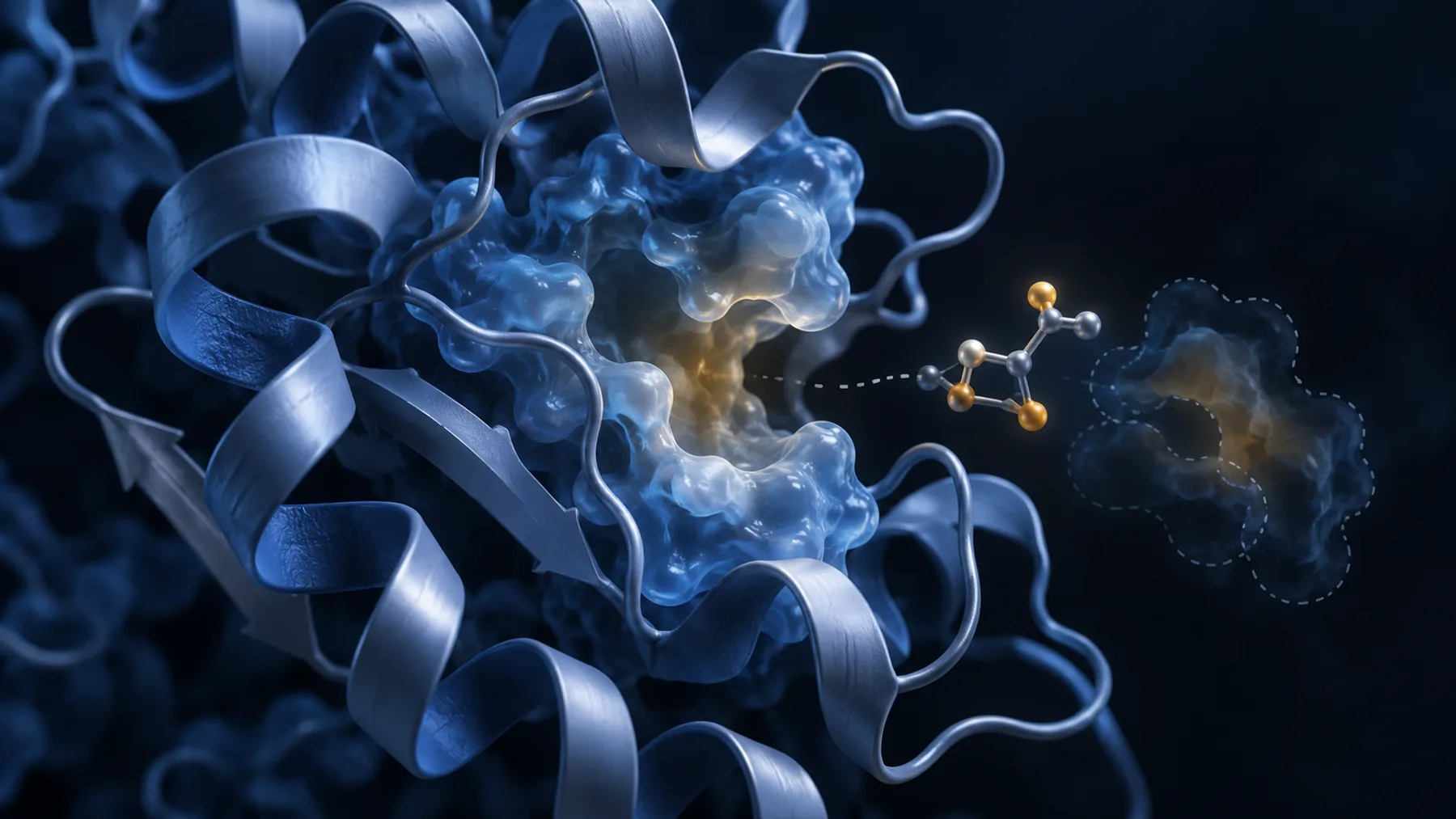
Intrinsically disordered proteins (IDPs) and regions (IDRs) do not populate one stable tertiary structure. They occupy ensembles whose dimensions and transient contacts depend on sequence, charge, PTMs, ligands, partners, temperature, and concentration. That changes the rules for expression, purification, quantification, chromatography, and structural interpretation.
The goal is not to force an IDP into one fold. It is to produce a chemically defined, monomeric or intentionally assembled ensemble that preserves the biology you plan to measure.
Key Takeaways
- Low AlphaFold confidence can be biologically correct. Long low-pLDDT regions often indicate disorder or partner-dependent folding, but pLDDT alone cannot distinguish every cause.
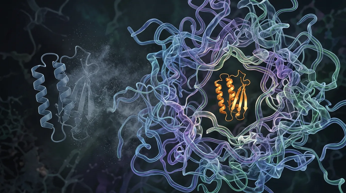
- AlphaFold returns coordinates; an IDP requires an ensemble. A single low-confidence model should not be interpreted as a native conformation, radius, or set of long-range contacts.
- Expect non-globular analytical behavior. IDPs often migrate anomalously on SDS-PAGE, elute early by SEC, and have weak A280 absorbance because of unusual charge, hydrodynamic size, and aromatic content.
- Proteolysis is often the first expression bottleneck. Use rapid cold handling, effective inhibitors, optimized termini, fusion strategies, or deliberately protected inclusion-body expression.
- Denaturing purification is not automatically destructive. For a protein that is natively disordered, urea-based capture followed by controlled buffer exchange can be cleaner than “native” purification through a heterogeneous lysate.
- Validate disorder with orthogonal methods. NMR, SAXS, CD, SEC-MALS, smFRET, and hydrodynamic measurements answer different ensemble questions.
Disorder Is a State, Not a Failed Fold
A globular protein typically hides hydrophobic residues in a compact core and occupies a narrow basin of conformations. An IDP is enriched in charged and polar residues, depleted in bulky hydrophobics, and lacks enough favorable intramolecular contacts to lock the chain into one tertiary structure under the condition of interest.
That does not mean “random coil” in the sense of no preferences. IDPs can contain:
- transient helices and β-structure;
- compact and expanded subensembles;
- molecular recognition features that fold upon partner binding;
- short linear motifs;
- PTM-regulated charge patterns;
- regions that drive phase separation;
- segments that become ordered in a complex.
The ensemble is the functional object. A buffer, mutation, tag, or purification step that compacts the chain may preserve solubility and destroy function.
Why AlphaFold Fails—and What the Failure Tells You
AlphaFold was trained to predict atomic structures represented in the Protein Data Bank. Its output is a small set of coordinate models. An IDP samples a distribution of structures, so no single coordinate set is the correct answer.
pLDDT is useful as a warning
In the human-proteome analysis, AlphaFold’s pLDDT separated experimentally resolved and unresolved residues and performed competitively on a disorder benchmark. The authors reported an area under the curve of 0.897 for pLDDT on the CAID DisProt-PDB set, while warning that low confidence can also mark regions structured only through interactions with other chains (Tunyasuvunakool et al., 2021).
That supports a careful interpretation:
- long pLDDT below 50 against a high-confidence core is evidence for disorder or context-dependent structure;
- a sharp boundary can help propose constructs;
- uniform low confidence across an entire chain may also reflect a shallow MSA, unusual composition, or lack of learned structural signal;
- a confident short helix inside an IDR may represent a transient or partner-bound motif—not a helix populated continuously in solution.
The coordinates are not an ensemble
Do not calculate a radius of gyration from the rank-1 AlphaFold IDP model and report it as a solution property. Do not treat transient contacts in a low-confidence tangle as binding sites. Do not dock a ligand into a pocket created by one arbitrary collapse of the chain.
Recent work illustrates the difference directly. A single AlphaFold model of TDP-43 disagreed with SAXS-derived distance distributions because its long disordered regions were represented by one conformation. An ensemble workflow constrained by AlphaFold-derived information and experimental data produced much better agreement (Sala et al., 2025).
AlphaFold can contribute restraints or identify folded islands. It does not eliminate ensemble measurement.
Partner-induced order is a separate prediction problem
If an IDR folds on binding, model the complex and compare apo/partner-bound evidence. High confidence in the bound helix does not prove that the free chain is helical. Low confidence in the apo monomer does not prove the region is functionless.
Our earlier article on why intrinsically disordered regions break protein pipelines focuses on construct design for mixed folded/disordered proteins. This guide covers the case where the disorder itself is the target.
Start by Defining the Biological Form
Before cloning, answer:
- Is the target a full-length IDP, one IDR from a larger protein, or a folded domain plus IDR?
- Which termini are native and functionally required?
- Are PTMs central to the experiment?
- Is a binding partner needed to protect or order the region?
- Is phase separation an intended property or a purification artifact to avoid?
- Does the assay require a completely native sequence, isotopic labeling, or an attachment site?
These choices determine whether you should use a removable fusion, a denaturing route, a eukaryotic host, or coexpression with a partner.
Expression Problem 1: Proteolysis
Extended solvent-exposed chains give proteases abundant access. IDPs also contain flexible termini and low-complexity segments that can create multiple cleavage products.
Diagnostic signature
- ladder of lower-molecular-weight bands;
- intact band immediately after lysis but rapid loss during purification;
- different fragments with N- and C-terminal antibodies;
- increased recovery at lower temperature or with faster handling.
First interventions
- harvest and lyse cold;
- use broad, compatible protease inhibition;
- shorten the time between lysis and capture;
- lower induction temperature and expression duration;
- move the affinity tag to the terminus you need to monitor;
- redesign a nonessential exposed terminus;
- test a protease-deficient strain;
- use a folded, removable fusion partner.
Confirm both ends. A His-tagged fragment can purify beautifully while most of the biological sequence is gone.
Expression Problem 2: Low Yield or Toxicity
Some IDPs interact promiscuously with nucleic acids, membranes, or cellular proteins. Others form condensates or expose degrons. High expression can therefore slow growth or select for deletion mutants.
First interventions
- reduce basal expression with a tighter vector/host;
- lower inducer and temperature;
- shorten induction;
- use autoinduction only after leakiness is controlled;
- express as a folded fusion such as MBP or SUMO;
- direct the fusion into inclusion bodies intentionally when soluble expression causes degradation or toxicity;
- move to cell-free or a eukaryotic host when PTMs or toxicity dominate.
Sequence-verify the expression plasmid after induction if escape mutants are plausible.
Fusion Tags: Protection Versus Perturbation
MBP, SUMO, GB1, thioredoxin, and other fusions can protect an IDP and improve recovery. They can also compact the chain, suppress phase behavior, create non-native contacts, or dominate hydrodynamic measurements.
Use a fusion as a production intermediate when possible:
His6–fusion–specific cleavage site–IDP
Then test cleavage at small scale and remove the tag immediately. SUMO is valuable when a native N-terminal residue is required. MBP provides stronger protection but a larger perturbation.
If the IDP precipitates after tag removal, do not assume it needs the tag to fold. Check pH, salt, concentration, oxidation, nucleic-acid contamination, and the intended biological assembly. Some IDPs become less soluble at high salt because electrostatic repulsion is screened; the rule can be opposite to a globular protein.
When Inclusion Bodies Are the Clean Route
For a natively disordered target, inclusion-body expression can protect the chain from proteolysis and simplify purification. The material is solubilized in urea or guanidinium, captured under denaturing conditions, cleaved if needed, and exchanged into the measurement buffer.
This is not conventional “refolding” toward one tertiary structure. It is removal of denaturant so the native ensemble can re-establish.
The Npro/EDDIE fusion strategy uses an autocleaving fusion that directs difficult peptides or IDPs into inclusion bodies. An optimized protocol solubilized the material in 8 M urea, purified it by IMAC, triggered cleavage during dialysis, removed much of the tag by precipitation or a second IMAC, and polished the IDP by reverse-phase HPLC (Tago et al., 2017).
Use this route when:
- soluble expression gives extensive proteolysis;
- isotopic labeling is required;
- the target tolerates denaturant and buffer exchange;
- the sequence lacks denaturant-sensitive covalent structure;
- you can verify the final ensemble and chemical integrity.
Heat and Acid: Exploit Them Carefully
Many IDPs remain soluble when globular E. coli proteins unfold and precipitate. Heat treatment can therefore be an effective early purification step.
Sic1, an intrinsically disordered cyclin-dependent-kinase inhibitor, was enriched by heating crude extract near boiling and removing precipitated host proteins. CD and mass spectrometry supported reversible behavior of the target under the tested conditions (Brocca et al., 2009). Dehydrin purifications have similarly used controlled heat precipitation to remove globular contaminants.
Do not generalize from those successes. Avoid heat when the IDP:
- contains heat-labile PTMs or covalent modifications;
- undergoes irreversible oxidation;
- forms β-rich aggregates or amyloid;
- has a folded partner/domain that must remain intact;
- changes its biological ensemble under heat in a way that does not reverse.
Run a microscale temperature series and validate recovery by mass, CD/NMR, and function.
Acid precipitation can also remove many contaminants while some acidic IDPs remain soluble. The same target-specific validation applies.
A Practical Purification Workflow
Step 1: Keep a complete mass balance
Save total lysate, soluble fraction, pellet, flow-through, wash, elution, cleavage, and polishing fractions. IDPs can disappear through proteolysis, nonspecific resin binding, adsorption to filters, or precipitation that is invisible at small scale.
Step 2: Remove nucleic acids early
Basic IDPs often bind DNA/RNA and produce high viscosity, abnormal A260/A280, and heterogeneous SEC. Use nuclease plus magnesium where compatible, high-salt washes, heparin chromatography, or ion exchange. Confirm that the treatment does not remove a biologically required nucleic acid.
Step 3: Capture under the condition that preserves identity
Use native IMAC for a stable soluble fusion, or denaturing IMAC when proteolysis/heterogeneity dominates. For an untagged acidic IDP, ion exchange can be powerful because the charge distribution differs from most host proteins.
Step 4: Remove the tag at low concentration
Test multiple target concentrations. IDP phase behavior can be sharply concentration dependent. Separate the released tag and protease promptly.
Step 5: Polish by chemistry, not apparent size alone
SEC is useful for aggregates and buffer exchange but misleading for molecular weight. Combine it with ion exchange, reverse-phase HPLC for compatible peptides/IDPs, or affinity depletion.
Step 6: Confirm chemical homogeneity
Use intact mass and, where needed, peptide mapping. IDPs are susceptible to oxidation, deamidation, phosphorylation loss, truncation, and heterogeneous initiator-methionine processing.
Step 7: Confirm the ensemble in the final buffer
The final buffer is part of the sample definition. Record pH, ionic strength, temperature, redox state, concentration, crowding agents, and binding partners.
Why Standard QC Lies to You
SDS-PAGE apparent mass
IDPs can bind SDS atypically and migrate according to charge/composition rather than calculated mass. Use intact mass to establish identity.
SEC apparent mass
An expanded chain has a large hydrodynamic radius and elutes earlier than a globular protein of the same mass. SEC-MALS separates hydrodynamic behavior from absolute molar mass more effectively.
A280 concentration
Many IDPs contain few tryptophan and tyrosine residues. Their extinction coefficient can be very low, making A280 noisy and highly sensitive to nucleic-acid contamination. Use amino-acid analysis, quantitative NMR, refractive index, a validated colorimetric assay, or absorbance at another wavelength where appropriate.
DLS polydispersity
A broad size signal may reflect an ensemble, aggregates, or both. DLS alone cannot distinguish them. Filter/centrifuge controls and SEC-MALS/SAXS provide stronger interpretation.
Choose the Structural Method for the Question
| Question | Primary method | What it tells you |
|---|---|---|
| Is the chain disordered? | CD + NMR | secondary-structure bias and residue-level dynamics |
| What are the ensemble dimensions? | SAXS, SEC-MALS, diffusion NMR | radius and hydrodynamic behavior |
| Are there transient contacts? | NMR PRE, smFRET, cross-linking with ensemble modeling | distance distributions and populations |
| Does a motif fold on binding? | NMR, CD, HDX-MS, complex structure | disorder-to-order transition |
| Does the protein phase separate? | microscopy, turbidity, centrifugation, concentration/salt series | coexistence and material behavior |
| Which residues drive the transition? | mutational scan with ensemble/phase readouts | sequence grammar of the behavior |
No single technique reconstructs the ensemble. Combine methods that constrain different observables.
Troubleshooting Decision Tree
No intact expression
Reduce leakiness and induction; use a protective fusion; test inclusion-body-directed expression; consider a different host if PTMs are essential.
Many fragments
Map cleavage sites; speed capture; move the tag; lower temperature; remove nonessential terminal degrons; use a denaturing route.
Soluble but sticky/nucleic-acid contaminated
Add nuclease, high-salt or heparin steps; compare pH and charge; test whether binding is biological.
Clean by gel but heterogeneous by SEC
Confirm intact mass; separate expanded monomer from oligomer with SEC-MALS, AUC, or native MS; run concentration and salt series.
Precipitates after tag removal
Lower concentration, change pH/salt, add the biological partner, check oxidation, and test whether the construct excludes a stabilizing neighboring region.
AlphaFold shows a confident island inside disorder
Predict the island alone and with known partners; inspect PAE and conservation; test it by NMR/CD or a binding-dependent construct. Do not assume permanent structure.
Worked Example: The “Oversized” IDP
An IDP calculated at 22 kDa runs near 32 kDa on SDS-PAGE and elutes at the position of a 60 kDa globular standard. The initial conclusion is dimerization.
A correct diagnosis uses:
- intact mass: 22 kDa;
- SEC-MALS: approximately monomeric molar mass across the peak;
- CD: low helicity, consistent with disorder;
- concentration series: no new high-mass species;
- NMR diffusion/SAXS: expanded hydrodynamic dimensions.
The sample was not an oligomer. Two globular-protein calibrations were being applied to a non-globular chain.
The Economics of an IDP Workflow
| Early decision | Cheap check | Expensive error avoided |
|---|---|---|
| Native vs denaturing purification | proteolysis time course | weeks optimizing unstable “native” lysate |
| Fusion retention/removal | microscale cleavage | liters of tag-dependent material |
| Monomer vs expanded chain | SEC-MALS/intact mass | unnecessary oligomer-disruption campaign |
| Single structure vs ensemble | CD/SAXS/NMR pilot | meaningless docking or mutation design |
| Disorder required vs removable | functional domain/IDR constructs | deleting the biology to improve crystallization |
IDPs are not inherently harder at every step. Some tolerate heat, denaturant, and acid better than globular proteins. The cost comes from using the wrong mental model.
Bottom Line
Treat an intrinsically disordered protein as an ensemble whose chemistry and dimensions depend on its environment. Use AlphaFold’s low confidence as a warning and a boundary clue, not as a structure. Design expression around proteolysis and toxicity, choose native or denaturing purification deliberately, and validate identity, absolute mass, and ensemble behavior with orthogonal methods.
The goal is not to make the protein look globular. It is to preserve the disordered state that carries the function.
Working With Disorder in Orbion
Orbion’s AstraUNFOLD provides per-residue disorder and amyloidogenicity predictions alongside transmembrane topology, while AlphaFold pLDDT and PAE tracks identify folded islands, uncertain boundaries, and context-dependent regions. Design lets you compare full-length, truncated, fusion, and partner-aware constructs without treating low pLDDT as an automatic deletion instruction. Bench then generates expression and purification plans with explicit QC gates for identity, cleavage, aggregation, and the intended assay.
For IDPs, the most useful prediction is often not a coordinate. It is a map of which experimental assumptions are unsafe.
References
- Tunyasuvunakool, K. et al. Highly accurate protein structure prediction for the human proteome. Nature 596, 590–596 (2021). doi:10.1038/s41586-021-03828-1
- Sala, D. et al. AlphaFold prediction of structural ensembles of disordered proteins. Nature Communications 16, 1837 (2025). doi:10.1038/s41467-025-56572-9
- Tago, K. et al. An optimized Npro-based method for the expression and purification of intrinsically disordered proteins for an NMR study. Protein Expression and Purification 134, 42–49 (2017). Full text
- Brocca, S. et al. Order propensity of an intrinsically disordered protein, the cyclin-dependent-kinase inhibitor Sic1. Proteins 76, 731–746 (2009). Full text
- Minde, D. P. et al. Large extent of disorder in Adenomatous Polyposis Coli offers a strategy to guard Wnt signalling against point mutations. PLoS ONE 8, e77257 (2013). doi:10.1371/journal.pone.0077257
- Marsh, J. A. & Forman-Kay, J. D. Sequence determinants of compaction in intrinsically disordered proteins. Biophysical Journal 98, 2383–2390 (2010). doi:10.1016/j.bpj.2010.02.006