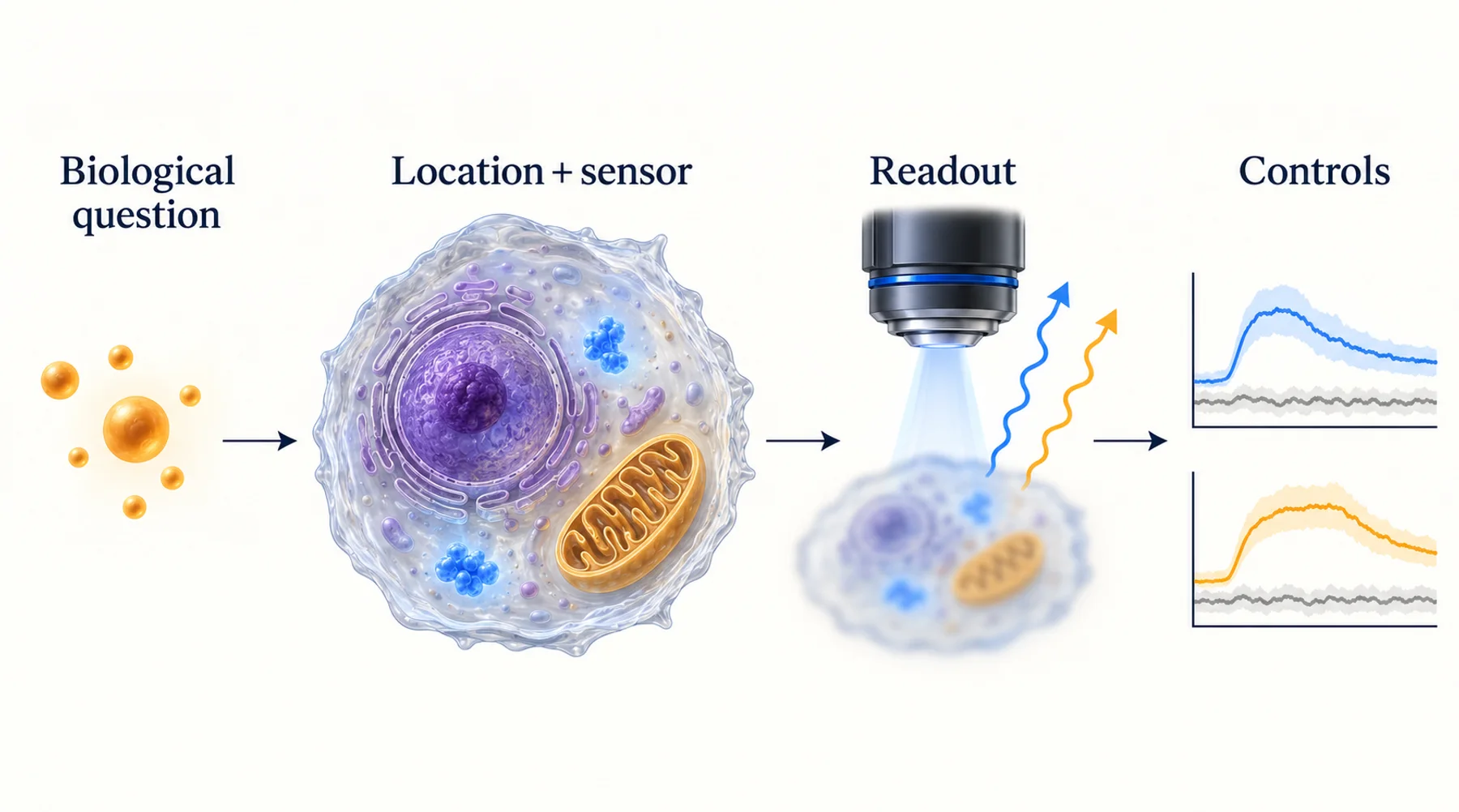
A treatment increases fluorescence in an oxidative-stress assay. It is tempting to report that “ROS increased.” But the probe may respond to a particular oxidation reaction, a redox relay, altered loading, pH, or the sample's own fluorescence. Those possibilities lead to different biological conclusions.
Choose a redox measurement by naming the chemical quantity or process the probe actually reports. Hydrogen peroxide, glutathione redox potential, and metabolic autofluorescence are related to cellular redox biology, but they are not interchangeable readouts of one universal oxidative-stress variable.
The practical goal is to connect probe chemistry to an interpretable cellular experiment. That requires controls for both the response mechanism and the environment in which the reporter operates.
Key Takeaways
- “ROS” covers chemically distinct species; a general fluorescence increase rarely identifies all of them.
- Peroxide reporters, glutathione-coupled reporters, and endogenous metabolic fluorescence answer different questions.
- A reversible redox sensor reflects oxidation and reduction processes acting on the reporter, not necessarily a simple analyte concentration.
- Check pH, localization, reporter abundance, illumination, and the usable response range.
- Use orthogonal evidence to test a biological interpretation without pretending that different assays measure the same quantity.
Replace “Oxidative Stress” With an Experimental Question
Oxidative stress can describe a biological condition involving altered oxidant production, antioxidant capacity, damage, or signaling. A fluorescence assay usually measures a narrower phenomenon. Start by deciding which of those phenomena matters to the experiment.
Do you need to follow a transient peroxide-associated response in the cytosol? Compare the glutathione redox environment of two compartments? Detect accumulated oxidative damage? Assess a metabolic response through endogenous fluorescence? Each question suggests different probes, time windows, and supporting measurements.
A treatment can change one readout without changing another in the same direction. A short signaling event may occur without detectable accumulated damage. A glutathione-coupled reporter may recover while an irreversible chemical product remains. A metabolic fluorescence signal can change because of several processes besides a change in peroxide.
Write the intended result in assay-specific language. “The cytosolic reporter ratio increased under these acquisition conditions” is an observation. “The treatment increased cytosolic peroxide concentration by a specified amount” requires additional calibration and mechanistic support. “The treatment caused oxidative damage” requires evidence for damage, not just a responsive reporter.
Three Readouts That Should Stay Separate
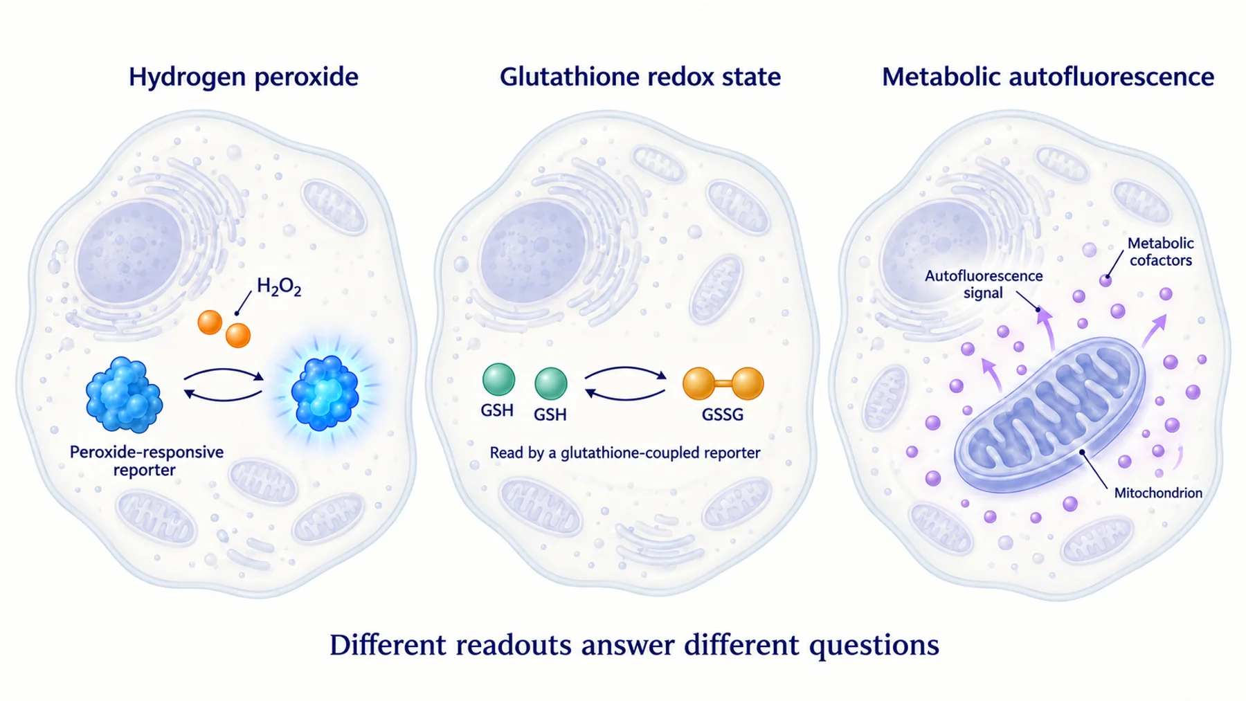
Figure 1. Different redox-related readouts have different response mechanisms. The pathways are conceptual and do not imply that one measurement can substitute directly for another.
Peroxide-Responsive Reporters
Genetically encoded peroxide reporters couple peroxide-sensitive chemistry to an optical output. HyPer7 is a published example designed for sensitive, ratiometric peroxide imaging. Its response should be interpreted with the controls and conditions relevant to the specific construct and cellular setting.
The important word is “peroxide,” not a generic “ROS.” Even a chemically well-characterized reporter operates in a cell that can change pH, reducing capacity, localization, and reporter integrity. Those influences belong in the experimental model.
Glutathione-Coupled Reporters
Grx1-roGFP2 couples a fluorescent redox reporter to glutathione chemistry through glutaredoxin. That is a different relationship from directly measuring the instantaneous concentration of hydrogen peroxide.
A glutathione-associated redox change can reflect several processes acting on the cellular redox system. It should not be relabelled as a peroxide concentration without a separate model and evidence. When reporting a derived redox potential, state the calibration, assumptions, and pH conditions needed for that calculation.
Metabolic Autofluorescence
Endogenous NAD(P)H- and flavin-associated signals can provide information about metabolic state. Label-free imaging of patient-derived organoids illustrates the use of these signals to assess treatment responses. Such measurements are valuable, but they are not a direct replacement for a chemically selective peroxide reporter.
The relative contributions of emitters, binding states, optics, and tissue structure matter. Define the exact optical index used rather than calling every ratio a “redox ratio” and assuming the same biological meaning across methods.
Understand the Oxidation–Reduction Balance
A reversible reporter can be driven toward an oxidized state by one process and returned toward a reduced state by another. The measured fraction or ratio therefore reflects the competition between those processes under the conditions of the experiment.
An illustrative two-state model writes the change in oxidized fraction x as dx/dt = kox(1 − x) − kred x. Here kox and kred are effective rates, not universal biochemical constants. At a steady state in this simplified model, x equals kox divided by kox + kred.
This shows why the same reporter oxidation can arise from increased oxidation, decreased reduction, or both. It also shows why recovery kinetics contain information different from the peak response. The model is a reasoning aid; actual reporters can involve additional states and reactions.
The roGFP2-Orp1 work provides a specific example of coupling a peroxidase-mediated oxidation relay to a fluorescent reporter. The lesson for assay design is to understand the relay, not to treat the fluorescence ratio as a universal peroxide meter.
If a treatment affects reducing systems as well as oxidant production, explicitly consider both. A sustained reporter response does not by itself distinguish persistent oxidant production from impaired recovery. Use targeted perturbations and independent observations to test those alternatives.
Ratiometric Does Not Mean Confounder-Free
Ratiometric reporters use two optical measurements to reduce some sources of variation. Depending on the design, they may involve two excitation bands, two emission bands, or energy transfer between fluorophores. The correction benefits depend on the underlying mechanism.
Both channels can still be affected by background, unequal bleaching, motion, saturation, or changes in the optical path. If the denominator is weak, the ratio can become unstable. Inspect the individual channels as well as the derived ratio, and retain them in the analysis record.
pH deserves specific attention because many fluorescent systems have pH-dependent behavior and biological treatments can alter pH. A reporter described as comparatively pH-stable still needs to be evaluated over the relevant conditions. Do not assume that a result for one member of a sensor family applies unchanged to another variant.
Where a response-deficient or pH-control construct is available, characterize its brightness and localization rather than assuming it is an otherwise identical control. A mutation can change folding or the reporter environment. The best control is one whose remaining differences are understood.
Chemical Probes Need Chemical Interpretation
Small-molecule probes can be practical when genetic modification is difficult or a short assay is sufficient. Their interpretation depends on uptake, intracellular conversion, reaction pathways, retention, efflux, and the optical properties of the products.
The methodological position paper by Kalyanaraman et al. explains why widely used oxidant-sensitive probes can support claims narrower than “total ROS.” Product identity and reaction specificity matter. A fluorescence signal alone may not distinguish the products or mechanisms needed for a species-specific conclusion.
Match the readout to the chemistry. If the probe accumulates an irreversible product, its time course can reflect cumulative reaction and product handling rather than the instantaneous concentration of the initiating species. If loading differs between conditions, a brighter population may contain more probe before any biological response occurs.
Include probe-free treated samples when the compound or cellular response might contribute fluorescence. Compare appropriate loading and retention controls. When the scientific claim depends on identifying a particular reaction product, consider an analytical method capable of resolving that product rather than relying only on a broad optical channel.
Design a Control Matrix Before Collecting the Main Dataset
The controls should separate competing explanations rather than merely produce a large positive response. A useful matrix includes reporter presence, treatment, illumination, and plausible nuisance variables.
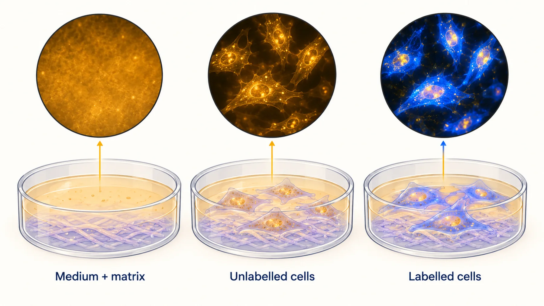
Figure 2. Each control tests a different alternative explanation. The control map is a planning framework; actual perturbations and acceptance criteria must be appropriate for the reporter and biological model.
| Alternative explanation | Useful comparison | What it can establish |
|---|---|---|
| Treatment changes autofluorescence | Treated unlabelled samples | Reporter-independent optical contribution |
| Reporter is outside its usable range | Suitable range or endpoint controls | Whether the assay retains response capacity |
| pH contributes to the signal | Validated pH measurement or control strategy | Whether pH changes could explain the observation |
| Imaging induces the response | Reduced-light or minimally observed samples | Dependence on the observation schedule |
| Reporter abundance changes biology | Controlled expression comparison | Whether the response depends on reporter burden |
| Signal reflects a damaged fusion | Reporter integrity and localization checks | Whether the intended sensor remains present |
No row proves the entire interpretation. A positive range control shows that the system can respond, but not necessarily that the treatment acts through the same mechanism. An unlabelled control reveals background, but not whether the reporter itself perturbs the biology.
Choose controls that are feasible and decisive for the claim. A small set of well-matched comparisons is usually more useful than a long list collected under different conditions. Predefine how the results will change the interpretation.
Compartment Targeting Changes the Meaning of the Result
The cytosol, mitochondrial matrix, intermembrane space, nucleus, and other compartments have distinct environments. A reporter targeted to one compartment does not automatically measure another, even if the compartments are close in a microscope image.
Check targeting, fusion orientation, and the integrity of the expressed construct. An optical pattern consistent with mitochondria can still leave uncertainty about the side of a membrane or the fraction of reporter outside the organelle. Use independent localization evidence appropriate to the resolution required by the claim.
Avoid comparing raw intensity across compartments as though it directly represented different analyte concentrations. Reporter abundance, pH, optical collection, and local chemistry can all differ. Establish the response range and calibration strategy separately where necessary.
Our article on tag placement provides useful background on how fusion architecture can alter protein behavior. In a sensor, that architecture can change both delivery and the relationship between the recognition element and the optical output.
A Worked Interpretation Example
Consider a hypothetical experiment in which a mitochondrial peroxide reporter shows a sustained ratio increase after treatment, while a cytosolic glutathione-coupled reporter changes only briefly. The two observations are not automatically contradictory.
They concern different chemistry in different compartments. The first step is to confirm that both sensors remain responsive and appropriately localized, and that the individual optical channels support the derived ratios. Check whether treatment changes background or pH enough to affect either result.
Next, examine recovery. If the peroxide-associated reporter remains oxidized after treatment removal, the explanation could involve continued oxidation, altered reduction, or reporter damage. A separate functional measure and a perturbation directed at the proposed mechanism can help distinguish these possibilities.
Finally, report the supported statement at the appropriate level. The experiment may show distinct reporter responses across compartments. It does not automatically quantify total cellular oxidant burden or establish which process caused a later phenotype. A careful interpretation preserves the useful observation while identifying the next discriminating experiment.
Keep Illumination Out of the Causal Story
Light can participate in the biology of a redox experiment. Repeated excitation may affect endogenous chromophores, reporter chemistry, and cellular function. If the treatment group requires more exposure because it is dimmer, the groups may receive different interventions during observation.
Use matched acquisition settings where that is appropriate, and document any differences. Compare a lower-exposure schedule and assess whether the same biological conclusion survives. A phototoxicity framework should be tailored to the cell type and process rather than reduced to a universal safe laser percentage.
Do not use an antioxidant merely to improve image stability without considering the question. In a redox assay, changing antioxidant capacity can alter the process being measured. Any such additive becomes part of the experimental condition and needs an explicit rationale and control.
Decide What the Data Can Support
Use relative units when calibration supports relative changes only. Report reporter oxidation or a defined ratio when that is the direct result. Convert to a concentration or potential only with a justified relationship, relevant calibration, and uncertainty.
State how cells were selected, how background was estimated, and how saturated or low-signal observations were handled. Show whether the result is consistent across independent preparations rather than relying on a large number of pixels from one sample.
For new protein sensors, the path from optical response to a redox application includes chemical selectivity, cellular reduction pathways, targeting, and validation against established approaches. Nebula is exploring responsive proteins as research tools; those application-specific properties remain to be demonstrated for its candidates.
Common Questions
Can one probe measure total ROS?
That phrase is usually broader than the chemistry supports. Identify the species, reaction, or response pathway measured by the probe and use additional assays for broader claims.
Does a higher reporter ratio mean more oxidative damage?
Not necessarily. A reporter response can reflect signaling or a reversible redox shift. Damage requires an appropriate damage endpoint and a design linking it to the observed process.
Should two redox assays agree numerically?
Only if they measure the same quantity under comparable conditions and calibrations. Different compartments, response kinetics, and chemistry can produce different results without either assay being wrong.
Diagram note: the cover and figures are conceptual illustrations, not experimental results. Numerical examples are identified in the text.