The first 25 residues are hydrophobic. One predictor calls them a cleavable signal peptide, another calls them transmembrane helix 1, and AlphaFold draws a confident helix with no membrane or cleavage event in sight. That disagreement changes the entire experiment: secreted protein versus membrane anchor, mature N terminus versus retained segment, soluble construct versus a topology-breaking truncation.
Signal peptides and N-terminal signal anchors share the feature that prediction tools see most clearly—a hydrophobic helix. The distinction depends on cleavage-site grammar, charge distribution, downstream topology, and the cellular machinery that recognizes the sequence. Resolve it by combining predictors that model both possibilities, then test processing and localization directly.
Key Takeaways
- A hydrophobic N terminus is not automatically a signal peptide. It may be a retained signal anchor or the first helix of a multipass membrane protein.
- Signal peptides have architecture, not one consensus sequence. An N-region, hydrophobic core, and cleavage-proximal C-region collectively support processing.
- Joint prediction reduces false assignments. Tools such as Phobius were designed to distinguish signal peptides from transmembrane segments rather than predicting them independently.
- Positive-inside charge is useful but not absolute. Flanking Lys/Arg residues help infer signal-anchor orientation, while topology can be altered by neighboring helices and folding.
- AlphaFold cannot show a proteolytic processing event. A modeled N-terminal helix may be a precursor feature that is absent from the mature protein.
- Validate cleavage and topology separately. Secretion, N-terminal mass, protease protection, glycosylation mapping, and membrane fractionation answer different questions.
Three N-Terminal Architectures That Look Similar in Sequence
Cleavable signal peptide
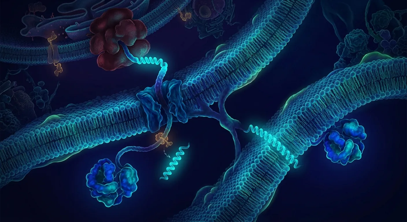
A classical secretory signal peptide directs the nascent chain to a translocation pathway and is cleaved by signal peptidase. It usually contains:
- a short N-region, often with positive residues;
- a hydrophobic H-region;
- a more polar C-region with cleavage-site preferences;
- a mature chain that begins immediately after cleavage.
Type I signal anchor
An N-terminal hydrophobic segment targets and anchors the protein but remains part of the mature chain. The N terminus is typically luminal/extracellular and the larger C-terminal region cytosolic or luminal depending on orientation and pathway context.
First helix of a multipass protein
The first hydrophobic segment participates in the final topology alongside later helices. Its orientation may depend on charge balance, neighboring segments, and cotranslational integration.
A fourth possibility is a mitochondrial or organellar targeting sequence, which can be amphipathic rather than strongly hydrophobic. Do not force every N-terminal targeting signal into the secretory-versus-TM binary.
Why Predictors Disagree
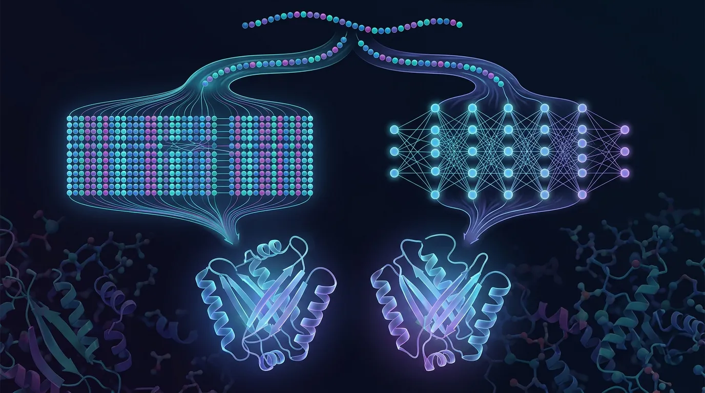
Independent signal-peptide and TM predictors can both score the same hydrophobic segment highly. A method trained only to find transmembrane helices may call the H-region a membrane span; a signal-peptide predictor may overread a hydrophobic signal anchor as cleavable.
Phobius addressed this directly with a joint hidden Markov model for signal peptides and transmembrane topology and improved discrimination between the two classes (Käll et al., 2004). Philius later used a dynamic Bayesian network to model topology and signal peptides from sequence (Reynolds et al., 2008). SignalP 6.0 uses protein language models and explicitly distinguishes several signal-peptide types (Teufel et al., 2022).
The useful workflow is not majority vote among ten correlated tools. Use one modern signal-peptide model, one joint topology model, and one independent topology model. Then inspect the sequence features that drive the disagreement.
Read the Sequence Around the Hydrophobic Core
Cleavage-site evidence
Signal peptidase I often favors small, neutral residues near positions −3 and −1 relative to cleavage. The broader C-region tends to be less hydrophobic than the core. A plausible cleavage site immediately after a strong hydrophobic segment supports a signal peptide.
The motif is probabilistic. A matching pair of small residues does not prove cleavage, and noncanonical sites occur.
Hydrophobic length and continuity
A long, uninterrupted hydrophobic segment extending through the proposed cleavage site supports a retained membrane helix. A signal peptide often shows a clearer transition from hydrophobic core to a polar cleavage region.
Flanking charge
Signal anchors often follow the positive-inside tendency: the side with more Lys/Arg residues is biased toward the cytosol. Classic signal-anchor experiments showed that charge substitutions flanking a hydrophobic segment can invert orientation (Sakaguchi et al., 1992).
This is a bias, not a law. Strong downstream domains, multiple helices, and translocon kinetics can modify topology.
Downstream topology
If the sequence contains several additional confident TM helices, the N-terminal segment is more likely part of a membrane topology—though a cleaved signal peptide can precede a multipass protein. Look for consistency across the whole chain.
What AlphaFold Can and Cannot Contribute
AlphaFold may model the hydrophobic segment as an α-helix with high pLDDT. Both signal peptides and transmembrane helices are helical, so that geometry does not distinguish them.
Three common interpretation errors follow:
- Treating the precursor as the mature protein. AlphaFold receives the full sequence and does not cleave the signal peptide.
- Inferring membrane retention from a helix. A cleaved signal peptide can be confidently helical before processing.
- Using packing in the model as proof. An N-terminal hydrophobic segment may pack artificially against the soluble domain when no membrane or translocon context is present.
Use pLDDT and PAE to judge the downstream folded domain and possible flexible junctions, not to infer cleavage. Our article on reading membrane topology from sequence covers full-chain topology reasoning.
A Practical Prediction Workflow
Step 1: Run a modern signal-peptide predictor
Record signal type, cleavage probability, and alternative cleavage positions—not just the binary label.
Step 2: Run a joint signal-peptide/TM model
Phobius or a comparable joint model asks the central discrimination question directly.
Step 3: Run whole-chain topology prediction
Count downstream helices, compare orientations, and identify whether excluding the N-terminal segment produces a coherent topology.
Step 4: Inspect the sequence manually
Mark N-region charge, hydrophobic-core boundaries, possible −3/−1 positions, prolines, polar interruptions, and charged residues on both flanks.
Step 5: Search homologs and experimental annotation
Conserved cleavage positions across close orthologs strengthen the signal-peptide case. Conserved hydrophobic length and flanking charge support an anchor. Treat automated database annotations as hypotheses unless backed by experiments.
Step 6: Design constructs that preserve both hypotheses
Do not commit to one interpretation prematurely. Build constructs that can report cleavage and localization.
The Minimal Experimental Panel
1. Full-length expression with both terminal readouts
Use N- and C-terminal antibodies or tags where they do not disrupt targeting. Loss of an N-terminal tag with retention of the C terminus supports processing, but the tag itself can alter recognition.
2. Membrane and soluble fractionation
Separate cytosol, membrane, and extracellular/periplasmic fractions. Include compartment-specific controls. A secreted protein can remain cell-associated, and a cleaved protein can still bind membranes peripherally.
3. N-terminal identity or intact mass
Mass spectrometry can establish whether the mature product lacks the predicted signal peptide and can locate heterogeneous cleavage.
4. Protease protection
In intact vesicles or microsomes, protease accessibility reports which domains face the protected lumen. Include detergent-permeabilized controls.
5. Glycosylation mapping
In a eukaryotic system, engineered or native glycosylation sites can report luminal exposure. Interpret only sites accessible to the glycosylation machinery.
6. Cleavage-site mutation
Change residues around the proposed cleavage site while preserving the hydrophobic core. Loss of processing without loss of membrane targeting supports a cleavable signal.
No single assay is enough. A soluble supernatant band might reflect lysis; membrane association might reflect aggregation; an N-terminal truncation might be host proteolysis.
Construct Design for the Two Hypotheses
If it is a cleavable signal peptide
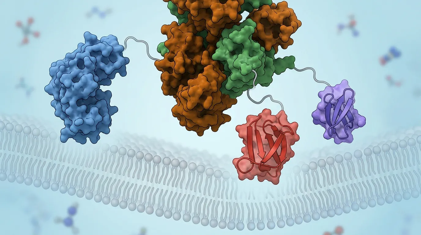
For secretion, keep the full native signal or replace it with a host-validated signal peptide. For cytosolic expression of the mature soluble domain, begin at the experimentally supported cleavage position and consider several nearby starts.
Our signal-peptide selection guide covers host-specific secretion design.
If it is a retained signal anchor
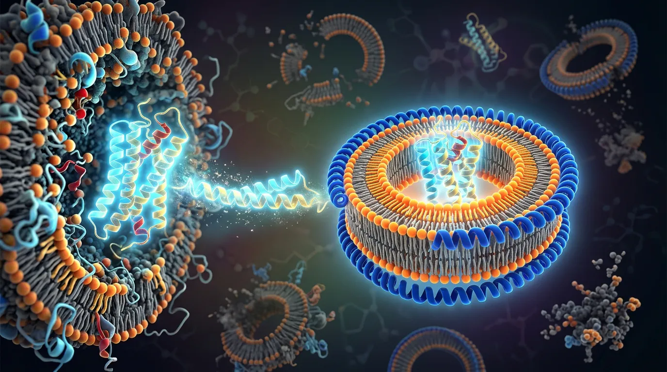
Removing the helix may destroy topology, stability, or oligomerization. Express the full-length membrane protein in a compatible host, screen detergents or reconstitution systems, and place tags on the experimentally accessible side.
If the call remains ambiguous
Build a compact panel:
- full-length precursor;
- mature-domain start at predicted cleavage site;
- ±2 and ±5 residue boundary variants;
- a cleavage-site-disrupting full-length mutant;
- optional hydrophobic-core mutant for localization control.
Compare expression, localization, processing, monodispersity, and function. The construct screen becomes the experiment that resolves the annotation.
The Expression Host Can Change the Answer
A signal peptide is interpreted by a particular translocation and signal-peptidase system. A sequence processed in mammalian cells may be inefficient in E. coli, yeast, insect cells, or a cell-free membrane system. Absence of cleavage in a heterologous host does not prove that the native protein has a retained anchor.
Host-dependent variables include:
- signal-recognition-particle specificity;
- cotranslational versus post-translational routing;
- signal-peptidase repertoire;
- membrane lipid composition;
- glycosylation and disulfide formation;
- translation rate and codon context;
- quality-control degradation.
When validating a biological annotation, use the native or a mechanistically similar host. When engineering secretion, judge the signal by the production host’s outcome. Those are different questions.
Bacterial Lipoprotein Signals Are a Separate Case
Some bacterial signal peptides are cleaved by signal peptidase II and contain a lipobox with a conserved cysteine that becomes lipidated. The mature protein remains membrane-associated through the lipid even though the peptide segment is removed.
A predictor that calls only “signal peptide” can therefore be technically right and experimentally misleading if you assume the product will be soluble. Check signal-peptide subtype, the residue immediately after the proposed cleavage, and the host’s lipidation machinery. SignalP 6.0 explicitly distinguishes several signal types, including lipoprotein signals.
For purification, a lipidated protein may require detergent despite cleavage. A mature construct beginning at the cysteine but expressed in the cytosol will not reproduce the native lipid anchor.
Tags Can Reverse the Outcome You Are Trying to Measure
An N-terminal His tag, fluorescent protein, or epitope can move positive charges, extend the N-region, obstruct signal-peptidase access, or prevent recognition entirely. A C-terminal tag is usually less disruptive for testing N-terminal processing, but it can still change topology if the C terminus contains retention or targeting information.
Use small tags and include an untagged or minimally tagged control. If detection requires an N-terminal tag, place a second readout downstream and verify that localization matches the native protein. Tag loss alone is not cleavage-site proof because host proteases can remove the tag.
Boundaries After Cleavage Are Often Heterogeneous
Signal peptidases can use nearby alternative sites. A single predicted position may represent a distribution, especially when the local sequence is suboptimal or overexpression saturates processing.
Map the mature N terminus directly and compare host, temperature, and expression level. For a soluble mature-domain construct, test starts at the dominant site and one or two neighboring residues. Boundary variants can differ sharply in solubility because the first residues may cap a helix, contribute a β-strand, or leave exposed hydrophobic sequence.
The correct construct start is the one that recreates the processed, folded product—not necessarily the highest-scoring computational cleavage coordinate.
Troubleshooting Decision Tree
SignalP says signal peptide; topology model says TM
Inspect cleavage score and flanking charge. Run a joint predictor and test N-terminal processing by mass spectrometry.
The full-length protein is in membrane fraction, but the mature construct is soluble
That supports either a cleavable signal peptide with inefficient secretion or a retained anchor. Determine whether the full-length product is cleaved.
The “mature” construct is insoluble
The chosen boundary may cut into the folded domain, or the N-terminal helix may be structurally required. Screen nearby starts and evaluate structure confidence/PAE at the junction.
The protein appears in culture medium
Rule out cell lysis with cytosolic markers. Confirm the N terminus and test whether secretion depends on the hydrophobic segment.
Protease protection gives mixed topology
The protein may insert in both orientations, vesicles may be leaky, or the sample may contain uninserted material. Add integrity controls and quantify rather than calling a single topology.
Homologs disagree
Closely related proteins can evolve different processing. Check alignment quality and compare the entire N-terminal architecture, not a single annotation field.
Worked Example: A 24-Residue Segment With Two Plausible Stories
A receptor-like protein has one N-terminal hydrophobic segment and a 300-residue enzymatic domain. SignalP predicts cleavage after residue 24; a TM predictor calls residues 5–25 a membrane helix.
The sequence shows small residues at −3/−1, a drop in hydrophobicity before residue 24, and few positive residues on the C-terminal flank. Joint prediction favors a signal peptide. Full-length expression yields a secreted product whose intact mass begins at residue 25. A cleavage-site mutant remains membrane-associated.
The initial TM call was not nonsensical—it detected the same hydrophobic targeting element. Processing data resolved its fate.
The Economics of Resolving the N Terminus Early
| Early decision | Cheap test | Expensive error avoided |
|---|---|---|
| cleaved vs retained | N-terminal mass | purifying the wrong molecular species |
| membrane vs secreted | fractionation controls | optimizing a host for an impossible compartment |
| correct mature boundary | small start-site panel | months on a destabilized truncation |
| topology orientation | protease protection | placing tags and domains on the wrong side |
| prediction disagreement | joint model + manual inspection | treating correlated votes as independent evidence |
Bottom Line
A hydrophobic N terminus tells you that the sequence may target a membrane; it does not tell you whether that segment survives in the mature protein. Combine signal-peptide and whole-chain topology models, inspect cleavage and charge grammar, build constructs that preserve both hypotheses, and validate processing and orientation with orthogonal experiments.
Resolving Topology in Orbion
Orbion Characterize places signal-peptide, transmembrane-topology, structure-confidence, disorder, and homology evidence in one target record. Design compares mature starts, full-length forms, boundary variants, and tag orientations. Bench generates a literature-grounded localization and processing workflow with the controls needed to distinguish secretion, insertion, and lysis.
The useful prediction is not merely “helix.” It is the next experiment that determines what the cell does with it.
References
- Teufel, F. et al. SignalP 6.0 predicts all five types of signal peptides using protein language models. Nature Biotechnology 40, 1023–1025 (2022). doi:10.1038/s41587-021-01156-3
- Käll, L., Krogh, A. and Sonnhammer, E. L. L. A combined transmembrane topology and signal peptide prediction method. Journal of Molecular Biology 338, 1027–1036 (2004). doi:10.1016/j.jmb.2004.03.016
- Reynolds, S. M., Käll, L., Riffle, M. E., Bilmes, J. A. and Noble, W. S. Transmembrane topology and signal peptide prediction using dynamic Bayesian networks. PLoS Computational Biology 4, e1000213 (2008). doi:10.1371/journal.pcbi.1000213
- Sakaguchi, M. et al. Functions of signal and signal-anchor sequences are determined by the balance between the hydrophobic segment and the N-terminal charge. Journal of Cell Biology 117, 1–11 (1992). doi:10.1083/jcb.117.1.15