You subcloned your antibody fragment — or your growth factor, or your protease inhibitor — into a clean pET vector, transformed BL21(DE3), induced, and ran a gel. The band is right there: strong, and at exactly the right molecular weight. Then one of two things happens. Either the supernatant is empty and your target is packed into inclusion bodies in the pellet. Or — sneakier, and worse — the protein is soluble, purifies cleanly, and is completely dead in your activity assay. Same root cause, and it has almost nothing to do with your IPTG concentration or your induction temperature.
The root cause is that your protein needs disulfide bonds to fold, and the E. coli cytoplasm is built to prevent exactly that. This guide is about why, and about the decision path out — periplasm, engineered strain, co-expressed foldase, cell-free, refold, or a different host — matched to your protein's biology instead of tried at random.
Key Takeaways
- The E. coli cytoplasm is actively reducing, by two independent pathways. The thioredoxin system and the glutathione/glutaredoxin system both keep cytoplasmic cysteines reduced — you have to disable both (the
trxBandgorgenes) to make the compartment oxidizing enough to form stable disulfides (Prinz et al., 1997). This is why "optimize the induction" never fixes it. - There are two distinct failure modes, and they point to different fixes. Inclusion bodies mean the chain never folded. Soluble but inactive usually means disulfides formed in the wrong pairing — a scrambled isomer. A non-reducing SDS-PAGE gel plus an activity assay tells them apart.
- Oxidation is not isomerization. Forming a disulfide is not the same as forming the right one. Proteins whose bonds are non-consecutive (interleaved along the chain) need a proofreading isomerase — DsbC or PDI — not just an oxidase. Get this distinction wrong and you ship soluble, scrambled, dead protein.
- Count cysteines first; it's free. An even cysteine count is consistent with N/2 disulfides; an odd count means at least one free (unpaired) cysteine you'll have to account for. An scFv carries two disulfides, a Fab five, an intact IgG more than a dozen — the higher the count and the more interleaved the topology, the more you need isomerization.
- Route by biology, not habit. Naturally secreted → lead with the periplasm. Non-consecutive bonds → a DsbC/PDI-equipped route. Glycosylated → a eukaryotic host, non-negotiable. Screening many constructs or the protein is toxic → cell-free with a defined redox buffer.
- There is no universal winner. The right route depends on bond count and topology, whether the protein is naturally secreted, glycosylation needs, and your throughput. Diagnose those four things up front and you pick one road instead of discovering which three don't work.
Disulfide-bonded proteins are not a niche problem. Most secreted and cell-surface proteins — most therapeutic targets, most antibodies and fragments, growth factors, cytokines, toxins, and hormones — carry disulfides that are load-bearing for the fold. And the most common reason they fail in standard E. coli is not the promoter, the codon usage, or the tag (Rosano & Ceccarelli, 2014). It's the redox chemistry of the compartment you asked them to fold in. The sections below build the mechanism, the menu of fixes, and the triage that chooses between them.
Why the E. coli Cytoplasm Refuses to Make Your Disulfides
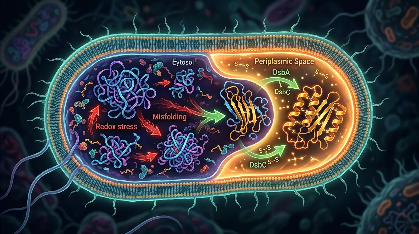
A disulfide bond is an oxidation: two cysteine thiols (–SH + HS–) lose electrons and form a covalent S–S bridge. That reaction only proceeds where the environment is oxidizing enough to pull those electrons away. The E. coli cytoplasm is the opposite of that — it is held in a strongly reducing state on purpose, because most cytoplasmic enzymes need their catalytic cysteines reduced to work.
Two independent enzymatic systems enforce that reducing state:
- The thioredoxin pathway — thioredoxin reductase (the
trxBgene product) uses NADPH to keep thioredoxins reduced, and reduced thioredoxin donates electrons to any disulfide that tries to form. - The glutathione/glutaredoxin pathway — glutathione reductase (the
gorgene product) keeps the cytoplasmic glutathione pool overwhelmingly in its reduced form (GSH), which in turn keeps the glutaredoxins reduced.
Knocking out one pathway isn't enough; the other compensates. You have to disable both — a trxB gor double mutant — before disulfide bonds accumulate in the cytoplasm (Prinz et al., 1997). Nature keeps the cytoplasm reduced for good reason, which is why disulfide-dependent proteins essentially never evolved to fold there (de Marco, 2009).
So when you overexpress a disulfide-dependent protein in a normal BL21 cytoplasm, its cysteines stay reduced. The chain has no way to lock in the bonds that stabilize its native fold, and one of two things happens:
- It aggregates into inclusion bodies. Without its disulfides, the folding intermediate exposes hydrophobic surface, collides with its neighbors, and precipitates (Baneyx & Mujacic, 2004). This is the "empty supernatant, fat band in the pellet" outcome. (If you're already staring at that pellet and disulfides may not be the cause, our companion piece on refold, redesign, or re-host walks the broader triage.)
- It stays soluble but never reaches the native state. Some proteins tolerate the reduced state and stay in solution as a loose, disulfide-free or partially scrambled conformation — a win on the gel and in the supernatant, right up until the activity assay reads zero.
That second outcome is the one that burns weeks, because it doesn't look like a folding failure. It looks like success. Which brings us to the distinction that decides your entire route.
Oxidation Is Not Isomerization: The Distinction That Decides Your Route
Here is the concept that most expression troubleshooting skips, and it is the most important one in this guide.
Making a disulfide bond is oxidation: join two cysteines. Making the correct set of disulfide bonds is a second, harder problem: isomerization — breaking wrong pairings and re-forming them until the native pattern wins.
Whether you need the second step depends on your protein's disulfide topology:
- Consecutive (nested or sequential) disulfides, where cysteines pair with nearby partners in a simple order, often form correctly with oxidation alone. The chain folds, the right cysteines end up adjacent, and a single oxidizing pass gets the native pattern.
- Non-consecutive (interleaved) disulfides, where cysteines pair with distant partners and the bonds cross over each other along the sequence, are a combinatorial trap. A fast oxidase will happily lock in the first pairing it encounters — often the wrong one. With n disulfides there are (2n−1)!! possible pairings; only one is native. Cystine-knot growth factors, many protease inhibitors, and complex secreted proteins live here.
For an interleaved topology, an oxidase that only forms bonds will give you soluble, fully-oxidized, scrambled protein: every cysteine bonded, none of them right, activity dead. You need a proofreader that can reshuffle mispaired bonds — an isomerase.
E. coli has both activities, but they live in the periplasm, split across four proteins (Messens & Collet, 2006):
- DsbA is the primary oxidase — a very oxidizing thioredoxin-fold protein that donates its own disulfide to a substrate cysteine pair, forming bonds rapidly and somewhat indiscriminately. Its discovery was the founding result of the whole field (Bardwell et al., 1991). DsbB re-oxidizes DsbA by passing electrons into the membrane respiratory chain.
- DsbC (with DsbD keeping it reduced) is the primary isomerase — it attacks incorrect disulfides and reshuffles them toward the native pattern.
The practical upshot for choosing a route: if your protein has multiple non-consecutive disulfides, every fix you pick must include an isomerase step. Periplasmic secretion gives you DsbC. SHuffle gives you a cytoplasmic DsbC. CyDisCo gives you PDI. An oxidase-only route — a bare trxB gor strain with no isomerase, or a cell-free reaction with only an oxidizing buffer — will scramble it. Half the "it expressed soluble but it's dead" stories are exactly this mismatch.
The Periplasm: E. coli's Built-In Oxidative Folding Compartment
The most robust place to fold a disulfide-bonded protein in E. coli is the compartment the bacterium already uses for its own disulfide-bonded proteins: the periplasm. It is oxidizing, and it carries the full Dsb machinery — DsbA/DsbB to oxidize, DsbC/DsbD to isomerize (Messens & Collet, 2006).
You get your protein there by fusing an N-terminal signal peptide that routes the nascent, unfolded chain through the Sec translocon into the periplasm, where it folds oxidatively. This is the standard, first-line move for antibody fragments (scFv, Fab) and small secreted proteins, and it's why so many antibody-discovery pipelines are built on periplasmic E. coli expression.
Three things to get right:
- The leader must match the host and the cargo. pelB and OmpA are the workhorse E. coli periplasmic leaders; PhoA is classic; the DsbA leader routes fast-folding chains co-translationally so they can't fold in the cytoplasm before export. Choosing among them is its own decision — secretion efficiency is a property of the leader paired with your protein — and we cover it in depth in signal peptide selection that actually works. Don't reuse whatever leader came with your vector.
- Isomerization may need a boost. For proteins with many non-consecutive bonds, native periplasmic DsbC can be limiting. Co-overexpressing DsbC in the periplasm measurably improves correct folding of complex-topology targets (Berkmen, 2012).
- The tradeoff is scale. The periplasm is small, with finite translocation capacity — yields are frequently lower than good cytoplasmic expression, and over-strong induction saturates the Sec machinery and backs product up as cytoplasmic inclusion bodies. It trades absolute titer for correct folding.
Use the periplasm when your protein is naturally secreted, needs isomerization, or is a standard antibody-fragment target. It's the highest-probability first attempt for most disulfide-rich proteins.
Engineered Oxidizing-Cytoplasm Strains: SHuffle, Origami, Rosetta-gami
If you want cytoplasmic yields — the larger compartment, the higher titers — but your protein needs disulfides, use a strain whose cytoplasm has been engineered to be oxidizing.
The foundational result: a trxB gor double mutant, missing both reductive pathways, allows disulfide bonds to form in the cytoplasm (Bessette et al., 1999). The commercial strains built on this idea:
- Origami / Rosetta-gami carry the
trxBandgormutations, making the cytoplasm oxidizing. They provide oxidation but no co-expressed isomerase by default — so they excel at proteins with simple/consecutive disulfides and can scramble proteins with interleaved topologies. (Rosetta-gami adds tRNAs for rare codons, orthogonal to the redox question.) - SHuffle goes a critical step further: it's a
trxB gorbackground that also constitutively expresses a cytoplasmic version of DsbC, so the cytoplasm both forms and proofreads disulfides — oxidation and isomerization in one strain (Lobstein et al., 2012). For a multi-disulfide, non-consecutive target, SHuffle is usually the better first choice than a bare Origami-type strain.
Two honest caveats. The trxB gor double knockout is metabolically costly — these strains grow more slowly and carry compensating mutations just to be viable, so plan for lower biomass. And an oxidizing cytoplasm is necessary but not sufficient: plenty of proteins still don't fold there. But for a disulfide-rich protein, a strain swap is frequently faster and higher-yielding than a refolding campaign — you change the host line, not the sequence.
CyDisCo and Co-Expressed Foldases: Oxidative Folding Without Rewiring the Strain
There's a third way to make disulfides in the cytoplasm that doesn't require knocking out the reducing pathways at all. Instead of removing the reductive machinery, you out-run it by co-expressing a dedicated oxidative-folding catalyst pair.
CyDisCo (Cytoplasmic Disulfide bond formation in E. coli) co-expresses two eukaryotic enzymes alongside your target: a sulfhydryl oxidase (Erv1p, from yeast mitochondria) that introduces disulfides, and a protein disulfide isomerase (PDI) that isomerizes them toward the native pattern. Introduced together, they fold multi-disulfide eukaryotic proteins in the cytoplasm of an otherwise normal (still-reducing) E. coli strain — the oxidase forms bonds fast enough, and PDI corrects them, before the thioredoxin/glutaredoxin systems can strip them back (Nguyen et al., 2011).
Why this matters as a distinct option: you keep your normal strain background (no trxB gor growth penalty, which helps biomass and yield); you get isomerization built in (PDI is a bona-fide isomerase, so CyDisCo handles non-consecutive topologies — it's not oxidase-only); and it scales, having been carried from shake flasks into fed-batch fermentation on defined media for higher-yield production (Gaciarz et al., 2017).
The cost is a co-expression plasmid to manage, plus the usual protein-dependent success rate. But when you want cytoplasmic folding and would rather not fight a sickly trxB gor strain, CyDisCo adds oxidative-folding capacity without re-plumbing the cell.
Cell-Free With a Defined Redox Buffer
Every route above happens inside a living cell, so the redox state is only as tunable as the cell's genetics allow. A cell-free reaction removes that constraint: it's an open tube, and you set the chemistry directly.
For disulfide work, you dial the glutathione redox buffer — the ratio of reduced (GSH) to oxidized (GSSG) glutathione — to whatever your protein wants, and add PDI or DsbC straight in as a soluble isomerase. Standard E. coli extract starts out reducing, so you treat it to stabilize an oxidizing potential and supply the foldases. Done right, this folds heavily bonded proteins that resist cellular routes: a plasminogen activator with nine disulfide bonds was folded in an E. coli cell-free system once the redox potential was stabilized and GSH/GSSG supplied (Yin & Swartz, 2004).
Use cell-free when you need precise redox control, fast screening of many constructs, or the protein is toxic to a living host. Don't when E. coli already works or you need grams — per-mg cost runs about an order of magnitude above a shake flask. The full cell-free-or-not decision, including lysate choice, is in when cell-free expression beats E. coli. The narrow point here: for a disulfide problem, cell-free's superpower is that redox and foldases become parameters you control, not host properties you're stuck with.
Refolding From Inclusion Bodies With a Redox Shuffle
If your protein went into inclusion bodies and you either need that exact sequence or expressed it insoluble on purpose (toxicity, maximum yield, purity), you can recover native protein in vitro. It's the lowest-throughput road, and the disulfide-specific step is the redox shuffle: after solubilizing the aggregate in strong chaotrope (6–8 M urea or 6 M guanidine) with a reducing agent to break scrambled bonds, you refold in a buffer of reduced and oxidized glutathione (GSH/GSSG) at a tuned ratio — the oxidized component drives bond formation, the reduced component lets wrong bonds break and re-form (isomerization in a tube). L-arginine (~0.4–0.5 M) suppresses aggregation during the process.
The honest reality: yields are protein-dependent and get worse as the disulfide count rises, because the space of wrong pairings grows and each screen must find the narrow window favoring the native pattern. A single-disulfide protein can refold well; a four- or nine-disulfide target often lands low or never folds. The full mechanics — dilution vs. dialysis vs. on-column, the additive screen — are in the inclusion-bodies decision guide. Reach for refolding when the sequence must stay fixed and the disulfide count is low, not as a reflex for every pellet.
When to Give Up on E. coli: Switch Host
Some disulfide-bonded proteins will never behave in E. coli, no matter which redox trick you deploy — because the missing ingredient isn't redox at all. Two triggers make a host switch non-negotiable:
- Glycosylation or other eukaryotic PTMs. E. coli cannot add N-linked glycans. If your protein's folding, stability, or function depends on glycosylation — as it does for most secreted human therapeutics — no strain, foldase, or refold rescues it. Full stop.
- Complex, many-disulfide folds that need the native chaperone stack. The eukaryotic ER is a purpose-built oxidative-folding factory — resident PDI family members, dedicated chaperones (BiP, calnexin/calreticulin), and quality control that retains misfolded chains. For proteins that evolved to fold there, reproducing it in a bacterium is often a losing battle.
Moving up the tree, yeast (Pichia pastoris, S. cerevisiae) offers secretion and some glycosylation cheaply; insect (baculovirus/Sf9) handles larger eukaryotic proteins and complexes; mammalian (HEK293, CHO) is the answer when human-like glycosylation and the full folding apparatus are required. The tradeoff is time and cost — mammalian is slowest and priciest. But for a glycosylated, disulfide-rich therapeutic, a host switch isn't the fallback; it's the correct first design decision.
The Fix Options at a Glance
| Route | How it makes native disulfides | Best for | Tradeoffs |
|---|---|---|---|
| Periplasmic secretion (Sec + Dsb) | Signal peptide exports the unfolded chain to the oxidizing periplasm; DsbA/DsbB oxidize, DsbC/DsbD isomerize | Naturally secreted proteins, antibody fragments, targets needing isomerization | Lower titer than cytoplasm; small compartment limits scale; leader/cargo matching required |
Origami / Rosetta-gami (trxB gor) | Oxidizing cytoplasm from double reductase knockout; oxidation only, no co-expressed isomerase | Simple/consecutive disulfides; cytoplasmic yield | Scrambles non-consecutive topologies; slower growth |
SHuffle (trxB gor + cytoplasmic DsbC) | Oxidizing cytoplasm plus a proofreading isomerase | Multiple/non-consecutive disulfides made cytoplasmically | Slower growth; still protein-dependent |
| CyDisCo (co-express Erv1p + PDI) | Sulfhydryl oxidase forms bonds + PDI isomerizes, out-running the reducing pathways in a normal strain | Cytoplasmic folding without a sick strain; eukaryotic multi-disulfide proteins | Extra co-expression plasmid; protein-dependent |
| Cell-free + defined redox buffer | Open reaction: set the GSH/GSSG ratio, add PDI/DsbC directly | Fast screening, toxic proteins, precise redox control | ~10× cost per mg; low throughput for production |
| Refold from inclusion bodies | Denature, then refold with a GSH/GSSG shuffle + arginine | You need this exact sequence; low disulfide count; high yield of denatured material | Low/variable recovery, worsens with more disulfides; weeks of screening |
| Switch host (insect / mammalian) | Native eukaryotic ER: PDI stack, chaperones, glycosylation | Glycosylated, many-disulfide, or natively-secreted-in-mammals proteins; therapeutics | Slower, costlier, more infrastructure |
The Triage: Five Questions Before You Clone
You don't need to try these routes in sequence and lose a month to each dead end. Five questions, answerable mostly from the sequence and the literature, route you before you order DNA.
1. How many cysteines, and how many expected disulfides? Count them. An even number is consistent with a fully bonded protein (N/2 bonds); an odd number means a free cysteine to account for (it can drive covalent aggregation if left exposed). More bonds means more difficulty and a stronger case for an isomerase-equipped route.
2. Is the protein naturally secreted? If its native localization is extracellular or cell-surface, it evolved to fold in an oxidizing compartment — so give it one. Secreted proteins are the strongest candidates for the periplasm and the worst for a bare reducing cytoplasm.
3. Are the disulfides consecutive or non-consecutive? The isomerization question from earlier. If the native bonds are interleaved (cystine-knot growth factors, complex inhibitors, multi-domain secreted proteins), you need a proofreading isomerase — periplasm-with-DsbC, SHuffle, CyDisCo, or a tuned refold — and should rule out oxidase-only routes like a bare Origami-type strain.
4. Is it also glycosylated (or dependent on other eukaryotic PTMs)? If yes, stop optimizing bacteria — glycosylation forces a eukaryotic host regardless of how the disulfide question resolves. It overrides everything else.
5. What's your scale and throughput? Screening many constructs or a toxic target → cell-free. Grams of a well-behaved secreted protein → periplasmic fermentation or a eukaryotic host. This exact sequence at any yield with few disulfides → refold. Match the route to the endpoint, not just the biochemistry.
Answer those five and usually one route stands out and two or three are clearly wrong. That's the game: spend bench time executing the likely winner, not eliminating the losers.
Decision Tree: Which Disulfide Route?
START: Disulfide-dependent protein misbehaving in BL21
        (inclusion bodies OR soluble-but-inactive)
│
├─ Also glycosylated / needs eukaryotic PTMs?
│     → SWITCH HOST (insect or mammalian). E. coli can't glycosylate — full stop.
│
├─ Naturally secreted protein?
│     → Lead with the PERIPLASM (pelB/OmpA/PhoA/DsbA leader).
│       It evolved to fold in an oxidizing compartment.
│
├─ Multiple, NON-CONSECUTIVE (interleaved) disulfides?
│     → You need ISOMERIZATION, not just oxidation. Choose one:
│         · Periplasm + co-expressed DsbC
│         · SHuffle (cytoplasmic DsbC proofreads)
│         · CyDisCo (Erv1p + PDI, normal strain)
│     → Do NOT use a bare Origami-type (oxidase-only) strain — it will scramble.
│
├─ Few / consecutive disulfides, want cytoplasmic yield?
│     → Origami / Rosetta-gami (oxidation is enough) OR SHuffle to be safe.
│
├─ Screening many constructs / protein is toxic / need tight redox control?
│     → CELL-FREE with a defined GSH:GSSG buffer + PDI.
│       [see the cell-free guide]
│
└─ Need THIS exact sequence, few disulfides, high yield of denatured OK?
      → REFOLD from inclusion bodies: solubilize (urea/GdnHCl) →
        GSH/GSSG redox shuffle + arginine → screen conditions.
        [see the inclusion-bodies guide]
Case Study: A Growth Factor That Expressed Soluble — and Dead
The following is an illustrative scenario, not a specific result — this failure mode is common enough to be worth walking through.
Problem. A team needed a small human growth factor — three disulfide bonds in a cystine-knot topology, non-consecutive connectivity — for a receptor-binding study. Knowing the E. coli cytoplasm is reducing, they were a step ahead of the usual mistake: they expressed in an Origami-type trxB gor strain to get cytoplasmic disulfides. And it worked — apparently. The protein was soluble at a respectable titer, purified cleanly, and ran as a tidy band. Then the binding assay came back flat. Repeatedly. No activity at all.
Analysis. Two observations reframed it. A non-reducing SDS-PAGE gel showed not one compact band but a smeary ladder — the signature of disulfide isomers, the same chain oxidized into several wrong bonding patterns. And the biology explained it: an Origami-type strain provides an oxidizing cytoplasm but no isomerase — it forms disulfides without proofreading them, and a cystine-knot fold is exactly the interleaved topology a fast oxidase traps in the wrong pairing. Every cysteine was bonded; almost none correctly. Soluble, fully oxidized, conformationally dead — the oxidation-without-isomerization failure, exactly.
Solution. The fix wasn't "form disulfides" — that was already happening — but "form the right ones," which meant adding an isomerase. The team ran two isomerase-equipped routes in parallel, sequence untouched: SHuffle, whose cytoplasmic DsbC reshuffles mispaired bonds toward the native set (Lobstein et al., 2012); and CyDisCo, co-expressing Erv1p and PDI so oxidation and isomerization run together in a healthier strain (Nguyen et al., 2011).
Outcome. SHuffle delivered a single compact non-reducing band and a live, dose-responsive assay; CyDisCo matched it. What rescued the project wasn't "can I make disulfides in the cytoplasm" — Origami already could — but "can I make the correct ones," which needs a proofreader. Dead assay to active protein: one expression round, because the failure mode named the missing ingredient.
Pre-Cloning Checklist
Before you order DNA for a disulfide-bonded target, verify:
- Cysteines counted — and expected disulfides estimated; any odd/unpaired cysteine flagged for handling.
- Native localization checked — is the protein naturally secreted or cell-surface (i.e., evolved to fold oxidizing)?
- Topology considered — consecutive vs. non-consecutive bonds; if interleaved, an isomerase-equipped route is mandatory.
- Glycosylation / eukaryotic-PTM need checked — if required, a eukaryotic host is non-negotiable and overrides the rest.
- Failure mode diagnosed — inclusion bodies (never folded) vs. soluble-but-inactive (scrambled), via a non-reducing gel plus an activity assay, not a reducing gel alone.
- Route matched to biology, not habit — isomerization-needing proteins routed to periplasm-with-DsbC, SHuffle, or CyDisCo — never a bare oxidase-only strain.
- Signal peptide chosen for the host if going periplasmic — matched to cargo, not carried over from the vector (see leader selection).
- Functional readout ready — so "soluble" is never mistaken for "folded and active."
The Economics: Cost per Route
The routes differ most in what a failure costs you, and in how quickly a failure announces itself. An oxidase-only strain fails silently — soluble, dead, discovered weeks later at the assay. A well-triaged route fails fast or not at all.
| Route | Setup effort | Time to folded protein | Yield profile | Where the cost hides |
|---|---|---|---|---|
| Periplasm | One construct (add a leader) | ~1 expression round | Moderate; periplasm-limited | Leader/cargo matching; lower titer |
| Origami / SHuffle | Strain swap (same plasmid) | ~1 expression round | Moderate–high when it works | Slow growth; Origami scrambles interleaved bonds |
| CyDisCo | Add a co-expression plasmid | ~1–2 rounds | Protein-dependent | Plasmid compatibility; still empirical |
| Cell-free | Reagents / kit | Hours per screen | mg-scale, expensive per mg | ~10× shake-flask cost per mg |
| Refold | Per-protein condition screen | Weeks | Low–moderate; drops sharply with more disulfides | Repeated screens; can fail outright on many-disulfide targets |
| Switch host | New vector + host | Weeks (mammalian longest) | High; native folding + glycosylation | Infrastructure, time, cost |
ROI consideration. The expensive mistake is almost never the reagents. It's spending three weeks refolding a four-disulfide protein SHuffle would have folded in one round — or worse, shipping soluble, oxidized, scrambled protein from an oxidase-only strain into a binding campaign and burning a month before someone runs a non-reducing gel. The five-question triage costs an afternoon; a silent isomerization failure costs a quarter. Decide the route from the biology first; spend bench time executing the one most likely to fold — correctly.
Bottom Line
Your disulfide-rich protein fails in standard E. coli because the cytoplasm is held reducing by two independent pathways, and disulfides can't survive there. The fix depends on four things: how many bonds and whether they're interleaved, whether the protein is naturally secreted, whether it's glycosylated, and your throughput. Naturally secreted → periplasm. Non-consecutive bonds → an isomerase-equipped route (periplasm+DsbC, SHuffle, or CyDisCo), never an oxidase-only strain. Glycosylated → a eukaryotic host, non-negotiable. Screening or toxic → cell-free with a tuned redox buffer. And remember the distinction that quietly kills projects: forming disulfides is not the same as forming the right ones. Diagnose before you clone.
How Orbion Helps
The entire triage above runs on signals you can read off the sequence and the protein's biology before you commit reagents — which is what Orbion is built to surface.
Start in Characterization. AstraSUIT returns experimental-suitability signals — classification, host association, membrane type, and subcellular location — so you can see whether your target reads as a secreted/periplasmic candidate, the strongest signal for the periplasmic route. AstraUNFOLD maps per-residue disorder, transmembrane topology, and amyloid/aggregation propensity, separating an aggregation-prone disulfide problem from a membrane-topology one that was never an inclusion-body question. And AstraPTM predicts residue-level PTMs with an expression-system filter showing which modifications a given host — E. coli, yeast, insect, mammalian — can actually make: the direct input to the glycosylation question, since a modification bacteria can't produce won't be rescued by any strain or refold. The sequence view shows your cysteine content directly.
One honest boundary: Orbion does not predict disulfide-bond connectivity. It won't give you the native pairing or classify your bonds as consecutive vs. non-consecutive — that comes from homology, literature, or experiment. What it gives you is the surrounding decision signal — secreted candidate?, aggregation risk?, host-compatible PTMs?, actually a membrane protein? You bring the connectivity; Orbion routes the rest.
When the signals point to a route, the Bench module lets you set the expression system explicitly — E. coli, insect, mammalian, or cell-free — and generates construct-aware protocols, with a Rate of Ease score (from solubility, disorder, aggregation, and topology signals) flagging predicted difficulty. When the answer is periplasmic secretion, the Design module's component library carries 14 curated signal peptides — including pelB, OmpA, PhoA, and DsbA for the E. coli periplasm and TorA for the Tat pathway — that assemble onto your construct in correct N→C order, codon-optimized for the target host (choosing among them).
Orbion won't pick your strain or run your fermentation — SHuffle vs. CyDisCo vs. periplasm is a bench call, and the wet lab always has the last mile. What it does is make sure you walk that mile down the right road, chosen from the protein's own signals, instead of discovering the reducing cytoplasm — or a scrambled-disulfide dead assay — three months in. Start at orbion.life.
References
-
Baneyx F, Mujacic M. (2004). Recombinant protein folding and misfolding in Escherichia coli. Nature Biotechnology, 22(11):1399–1408. https://doi.org/10.1038/nbt1029
-
Prinz WA, Åslund F, Holmgren A, Beckwith J. (1997). The role of the thioredoxin and glutaredoxin pathways in reducing protein disulfide bonds in the Escherichia coli cytoplasm. Journal of Biological Chemistry, 272(25):15661–15667. https://doi.org/10.1074/jbc.272.25.15661
-
Bardwell JCA, McGovern K, Beckwith J. (1991). Identification of a protein required for disulfide bond formation in vivo. Cell, 67(3):581–589. https://doi.org/10.1016/0092-8674(91)90532-4
-
Messens J, Collet JF. (2006). Pathways of disulfide bond formation in Escherichia coli. The International Journal of Biochemistry & Cell Biology, 38(7):1050–1062. https://doi.org/10.1016/j.biocel.2005.12.011
-
de Marco A. (2009). Strategies for successful recombinant expression of disulfide bond-dependent proteins in Escherichia coli. Microbial Cell Factories, 8:26. https://doi.org/10.1186/1475-2859-8-26
-
Berkmen M. (2012). Production of disulfide-bonded proteins in Escherichia coli. Protein Expression and Purification, 82(1):240–251. https://doi.org/10.1016/j.pep.2011.10.009
-
Bessette PH, Åslund F, Beckwith J, Georgiou G. (1999). Efficient folding of proteins with multiple disulfide bonds in the Escherichia coli cytoplasm. Proceedings of the National Academy of Sciences USA, 96(24):13703–13708. https://doi.org/10.1073/pnas.96.24.13703
-
Lobstein J, Emrich CA, Jeans C, Faulkner M, Riggs P, Berkmen M. (2012). SHuffle, a novel Escherichia coli protein expression strain capable of correctly folding disulfide bonded proteins in its cytoplasm. Microbial Cell Factories, 11:56. https://doi.org/10.1186/1475-2859-11-56
-
Nguyen VD, Hatahet F, Salo KEH, Enlund E, Zhang C, Ruddock LW. (2011). Pre-expression of a sulfhydryl oxidase significantly increases the yields of eukaryotic disulfide bond containing proteins expressed in the cytoplasm of E. coli. Microbial Cell Factories, 10:1. https://doi.org/10.1186/1475-2859-10-1
-
Gaciarz A, Khatri NK, Velez-Suberbie ML, Saaranen MJ, Uchida Y, Keshavarz-Moore E, Ruddock LW. (2017). Efficient soluble expression of disulfide bonded proteins in the cytoplasm of Escherichia coli in fed-batch fermentations on chemically defined minimal media. Microbial Cell Factories, 16:108. https://doi.org/10.1186/s12934-017-0721-x
-
Yin G, Swartz JR. (2004). Enhancing multiple disulfide bonded protein folding in a cell-free system. Biotechnology and Bioengineering, 86(2):188–195. https://doi.org/10.1002/bit.10827
-
Rosano GL, Ceccarelli EA. (2014). Recombinant protein expression in Escherichia coli: advances and challenges. Frontiers in Microbiology, 5:172. https://doi.org/10.3389/fmicb.2014.00172